Ruthenium »
PDB 6bo2-8oyr »
6fh8 »
Ruthenium in PDB 6fh8: E. Coli Surface Display of Streptavidin For Directed Evolution of An Allylic Deallocase
Protein crystallography data
The structure of E. Coli Surface Display of Streptavidin For Directed Evolution of An Allylic Deallocase, PDB code: 6fh8
was solved by
T.Heinisch,
F.Schwizer,
B.Garabedian,
E.Csibra,
M.Jeschek,
V.Pinheirobernhardes,
P.Marliere,
S.Panke,
T.R.Ward,
with X-Ray Crystallography technique. A brief refinement statistics is given in the table below:
| Resolution Low / High (Å) | 45.80 / 1.64 |
| Space group | I 41 2 2 |
| Cell size a, b, c (Å), α, β, γ (°) | 57.722, 57.722, 183.615, 90.00, 90.00, 90.00 |
| R / Rfree (%) | 12 / 15.9 |
Ruthenium Binding Sites:
The binding sites of Ruthenium atom in the E. Coli Surface Display of Streptavidin For Directed Evolution of An Allylic Deallocase
(pdb code 6fh8). This binding sites where shown within
5.0 Angstroms radius around Ruthenium atom.
In total only one binding site of Ruthenium was determined in the E. Coli Surface Display of Streptavidin For Directed Evolution of An Allylic Deallocase, PDB code: 6fh8:
In total only one binding site of Ruthenium was determined in the E. Coli Surface Display of Streptavidin For Directed Evolution of An Allylic Deallocase, PDB code: 6fh8:
Ruthenium binding site 1 out of 1 in 6fh8
Go back to
Ruthenium binding site 1 out
of 1 in the E. Coli Surface Display of Streptavidin For Directed Evolution of An Allylic Deallocase

Mono view
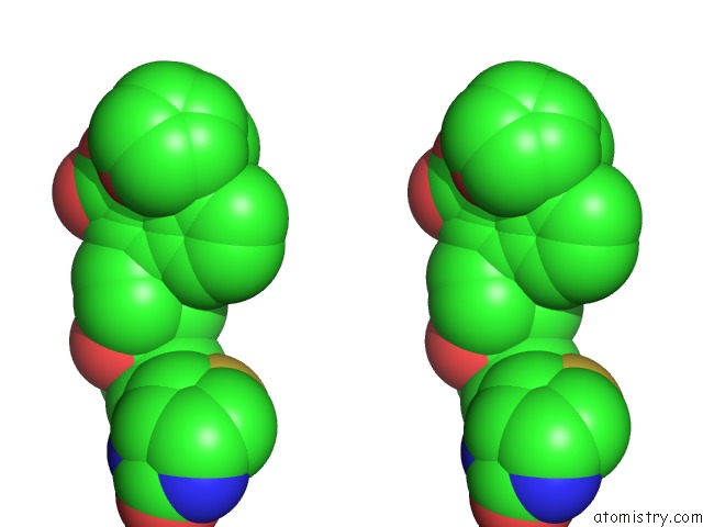
Stereo pair view
Mono view
Stereo pair view
A full contact list of Ruthenium with other atoms in the Ru binding
site number 1 of E. Coli Surface Display of Streptavidin For Directed Evolution of An Allylic Deallocase within 5.0Å range:
|
Reference:
T.Heinisch,
F.Schwizer,
B.Garabedian,
E.Csibra,
M.Jeschek,
J.Vallapurackal,
V.B.Pinheiro,
P.Marliere,
S.Panke,
T.R.Ward.
E. Colisurface Display of Streptavidin For Directed Evolution of An Allylic Deallylase. Chem Sci V. 9 5383 2018.
ISSN: ISSN 2041-6520
PubMed: 30079176
DOI: 10.1039/C8SC00484F
Page generated: Thu Oct 10 13:06:16 2024
ISSN: ISSN 2041-6520
PubMed: 30079176
DOI: 10.1039/C8SC00484F
Last articles
Fe in 2YXOFe in 2YRS
Fe in 2YXC
Fe in 2YNM
Fe in 2YVJ
Fe in 2YP1
Fe in 2YU2
Fe in 2YU1
Fe in 2YQB
Fe in 2YOO