Ruthenium »
PDB 4l55-5ira »
5ifo »
Ruthenium in PDB 5ifo: X-Ray Structure of Hsa-Myr-KP1019
Protein crystallography data
The structure of X-Ray Structure of Hsa-Myr-KP1019, PDB code: 5ifo
was solved by
A.Bijelic,
S.Theiner,
B.K.Keppler,
A.Rompel,
with X-Ray Crystallography technique. A brief refinement statistics is given in the table below:
| Resolution Low / High (Å) | 45.84 / 3.20 |
| Space group | C 1 2 1 |
| Cell size a, b, c (Å), α, β, γ (°) | 181.110, 38.060, 94.950, 90.00, 105.06, 90.00 |
| R / Rfree (%) | 24.4 / 26.2 |
Ruthenium Binding Sites:
The binding sites of Ruthenium atom in the X-Ray Structure of Hsa-Myr-KP1019
(pdb code 5ifo). This binding sites where shown within
5.0 Angstroms radius around Ruthenium atom.
In total 2 binding sites of Ruthenium where determined in the X-Ray Structure of Hsa-Myr-KP1019, PDB code: 5ifo:
Jump to Ruthenium binding site number: 1; 2;
In total 2 binding sites of Ruthenium where determined in the X-Ray Structure of Hsa-Myr-KP1019, PDB code: 5ifo:
Jump to Ruthenium binding site number: 1; 2;
Ruthenium binding site 1 out of 2 in 5ifo
Go back to
Ruthenium binding site 1 out
of 2 in the X-Ray Structure of Hsa-Myr-KP1019
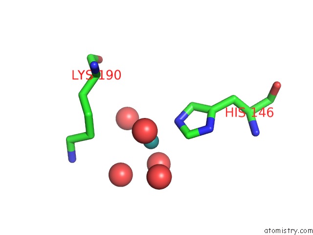
Mono view
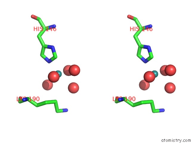
Stereo pair view
Mono view
Stereo pair view
A full contact list of Ruthenium with other atoms in the Ru binding
site number 1 of X-Ray Structure of Hsa-Myr-KP1019 within 5.0Å range:
|
Ruthenium binding site 2 out of 2 in 5ifo
Go back to
Ruthenium binding site 2 out
of 2 in the X-Ray Structure of Hsa-Myr-KP1019
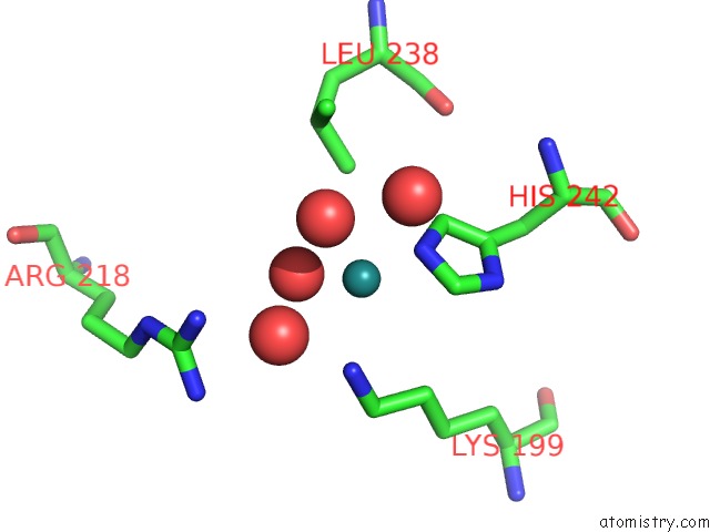
Mono view
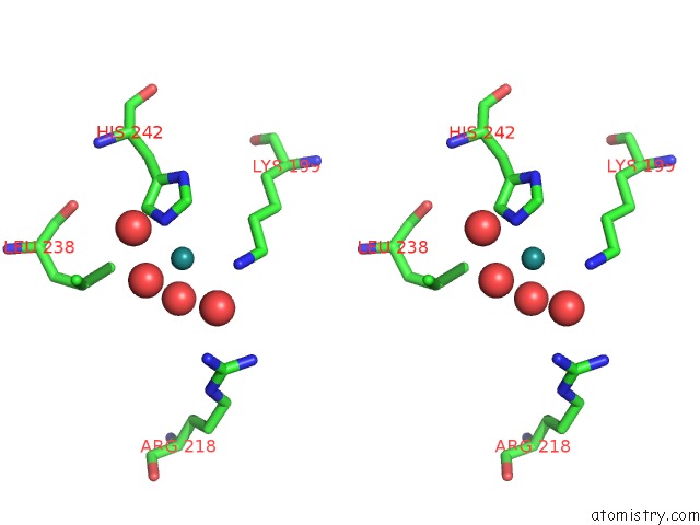
Stereo pair view
Mono view
Stereo pair view
A full contact list of Ruthenium with other atoms in the Ru binding
site number 2 of X-Ray Structure of Hsa-Myr-KP1019 within 5.0Å range:
|
Reference:
A.Bijelic,
S.Theiner,
B.K.Keppler,
A.Rompel.
X-Ray Structure Analysis of Indazolium Trans-[Tetrachlorobis(1H-Indazole)Ruthenate(III)] (KP1019) Bound to Human Serum Albumin Reveals Two Ruthenium Binding Sites and Provides Insights Into the Drug Binding Mechanism. J.Med.Chem. V. 59 5894 2016.
ISSN: ISSN 0022-2623
PubMed: 27196130
DOI: 10.1021/ACS.JMEDCHEM.6B00600
Page generated: Thu Oct 10 12:58:53 2024
ISSN: ISSN 0022-2623
PubMed: 27196130
DOI: 10.1021/ACS.JMEDCHEM.6B00600
Last articles
K in 3OSDK in 3OL5
K in 3NZ7
K in 3NYP
K in 3OIA
K in 3OD9
K in 3NR6
K in 3N25
K in 3NKR
K in 3NKQ