Ruthenium »
PDB 3m0b-4kgc »
3m0b »
Ruthenium in PDB 3m0b: Ru-Porphyrin Protein Scaffolds For Sensing O2
Protein crystallography data
The structure of Ru-Porphyrin Protein Scaffolds For Sensing O2, PDB code: 3m0b
was solved by
M.B.Winter,
E.J.Mclaurin,
S.Y.Reece,
C.Olea Jr.,
D.G.Nocera,
M.A.Marletta,
with X-Ray Crystallography technique. A brief refinement statistics is given in the table below:
| Resolution Low / High (Å) | 32.35 / 2.00 |
| Space group | P 61 2 2 |
| Cell size a, b, c (Å), α, β, γ (°) | 61.183, 61.183, 245.116, 90.00, 90.00, 120.00 |
| R / Rfree (%) | 20.4 / 22.5 |
Ruthenium Binding Sites:
The binding sites of Ruthenium atom in the Ru-Porphyrin Protein Scaffolds For Sensing O2
(pdb code 3m0b). This binding sites where shown within
5.0 Angstroms radius around Ruthenium atom.
In total only one binding site of Ruthenium was determined in the Ru-Porphyrin Protein Scaffolds For Sensing O2, PDB code: 3m0b:
In total only one binding site of Ruthenium was determined in the Ru-Porphyrin Protein Scaffolds For Sensing O2, PDB code: 3m0b:
Ruthenium binding site 1 out of 1 in 3m0b
Go back to
Ruthenium binding site 1 out
of 1 in the Ru-Porphyrin Protein Scaffolds For Sensing O2
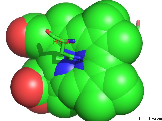
Mono view
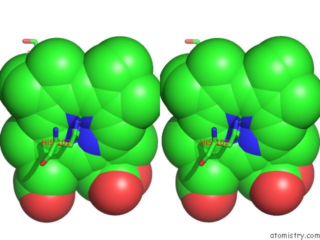
Stereo pair view
Mono view
Stereo pair view
A full contact list of Ruthenium with other atoms in the Ru binding
site number 1 of Ru-Porphyrin Protein Scaffolds For Sensing O2 within 5.0Å range:
|
Reference:
M.B.Winter,
E.J.Mclaurin,
S.Y.Reece,
C.Olea,
D.G.Nocera,
M.A.Marletta.
Ru-Porphyrin Protein Scaffolds For Sensing O2. J.Am.Chem.Soc. V. 132 5582 2010.
ISSN: ISSN 0002-7863
PubMed: 20373741
DOI: 10.1021/JA101527R
Page generated: Tue Aug 19 00:50:58 2025
ISSN: ISSN 0002-7863
PubMed: 20373741
DOI: 10.1021/JA101527R
Last articles
Mn in 9LJUMn in 9LJW
Mn in 9LJS
Mn in 9LJR
Mn in 9LJT
Mn in 9LJV
Mg in 9UA2
Mg in 9R96
Mg in 9VM1
Mg in 9P01