Ruthenium »
PDB 1bex-3fy0 »
3csf »
Ruthenium in PDB 3csf: Crystal Structure of PI3K P110GAMMA Catalytical Domain in Complex with Organoruthenium Inhibitor DW2
Enzymatic activity of Crystal Structure of PI3K P110GAMMA Catalytical Domain in Complex with Organoruthenium Inhibitor DW2
All present enzymatic activity of Crystal Structure of PI3K P110GAMMA Catalytical Domain in Complex with Organoruthenium Inhibitor DW2:
2.7.1.153;
2.7.1.153;
Protein crystallography data
The structure of Crystal Structure of PI3K P110GAMMA Catalytical Domain in Complex with Organoruthenium Inhibitor DW2, PDB code: 3csf
was solved by
P.Xie,
R.Marmorstein,
with X-Ray Crystallography technique. A brief refinement statistics is given in the table below:
| Resolution Low / High (Å) | 50.00 / 2.80 |
| Space group | C 1 2 1 |
| Cell size a, b, c (Å), α, β, γ (°) | 143.635, 68.082, 106.270, 90.00, 95.26, 90.00 |
| R / Rfree (%) | 25.2 / 28.7 |
Ruthenium Binding Sites:
The binding sites of Ruthenium atom in the Crystal Structure of PI3K P110GAMMA Catalytical Domain in Complex with Organoruthenium Inhibitor DW2
(pdb code 3csf). This binding sites where shown within
5.0 Angstroms radius around Ruthenium atom.
In total only one binding site of Ruthenium was determined in the Crystal Structure of PI3K P110GAMMA Catalytical Domain in Complex with Organoruthenium Inhibitor DW2, PDB code: 3csf:
In total only one binding site of Ruthenium was determined in the Crystal Structure of PI3K P110GAMMA Catalytical Domain in Complex with Organoruthenium Inhibitor DW2, PDB code: 3csf:
Ruthenium binding site 1 out of 1 in 3csf
Go back to
Ruthenium binding site 1 out
of 1 in the Crystal Structure of PI3K P110GAMMA Catalytical Domain in Complex with Organoruthenium Inhibitor DW2
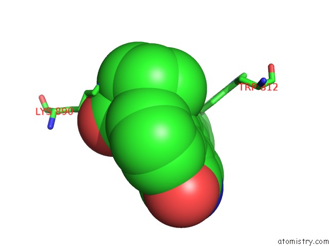
Mono view
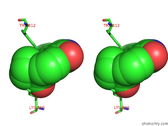
Stereo pair view
Mono view
Stereo pair view
A full contact list of Ruthenium with other atoms in the Ru binding
site number 1 of Crystal Structure of PI3K P110GAMMA Catalytical Domain in Complex with Organoruthenium Inhibitor DW2 within 5.0Å range:
|
Reference:
P.Xie,
D.S.Williams,
G.E.Atilla-Gokcumen,
L.Milk,
M.Xiao,
K.S.Smalley,
M.Herlyn,
E.Meggers,
R.Marmorstein.
Structure-Based Design of An Organoruthenium Phosphatidyl-Inositol-3-Kinase Inhibitor Reveals A Switch Governing Lipid Kinase Potency and Selectivity. Acs Chem.Biol. V. 3 305 2008.
ISSN: ISSN 1554-8929
PubMed: 18484710
DOI: 10.1021/CB800039Y
Page generated: Tue Aug 19 00:50:12 2025
ISSN: ISSN 1554-8929
PubMed: 18484710
DOI: 10.1021/CB800039Y
Last articles
Mn in 9LJUMn in 9LJW
Mn in 9LJS
Mn in 9LJR
Mn in 9LJT
Mn in 9LJV
Mg in 9UA2
Mg in 9R96
Mg in 9VM1
Mg in 9P01