Ruthenium »
PDB 1bex-3fy0 »
3ase »
Ruthenium in PDB 3ase: Crystal Structure of Zinc Myoglobin Soaked with RU3O Cluster
Protein crystallography data
The structure of Crystal Structure of Zinc Myoglobin Soaked with RU3O Cluster, PDB code: 3ase
was solved by
T.Koshiyama,
M.Shirai,
T.Hikage,
H.Tabe,
K.Tanaka,
S.Kitagawa,
T.Ueno,
with X-Ray Crystallography technique. A brief refinement statistics is given in the table below:
| Resolution Low / High (Å) | 19.65 / 1.75 |
| Space group | P 6 |
| Cell size a, b, c (Å), α, β, γ (°) | 90.722, 90.722, 45.395, 90.00, 90.00, 120.00 |
| R / Rfree (%) | 20.1 / 23.2 |
Other elements in 3ase:
The structure of Crystal Structure of Zinc Myoglobin Soaked with RU3O Cluster also contains other interesting chemical elements:
| Zinc | (Zn) | 1 atom |
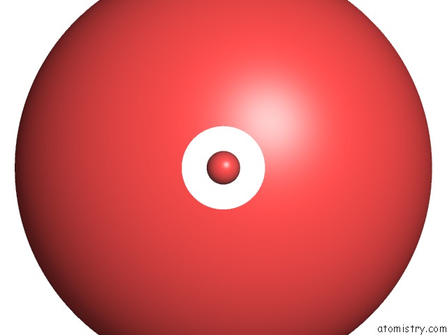
Ruthenium Binding Sites:
The binding sites of Ruthenium atom in the Crystal Structure of Zinc Myoglobin Soaked with RU3O Cluster
(pdb code 3ase). This binding sites where shown within
5.0 Angstroms radius around Ruthenium atom.
In total 6 binding sites of Ruthenium where determined in the Crystal Structure of Zinc Myoglobin Soaked with RU3O Cluster, PDB code: 3ase:
Jump to Ruthenium binding site number: 1; 2; 3; 4; 5; 6;
In total 6 binding sites of Ruthenium where determined in the Crystal Structure of Zinc Myoglobin Soaked with RU3O Cluster, PDB code: 3ase:
Jump to Ruthenium binding site number: 1; 2; 3; 4; 5; 6;
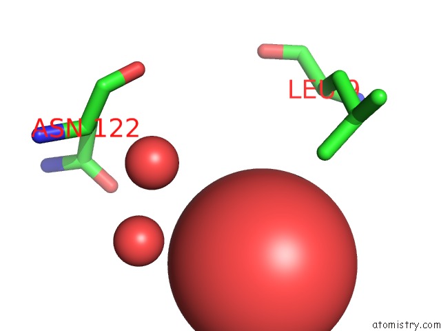
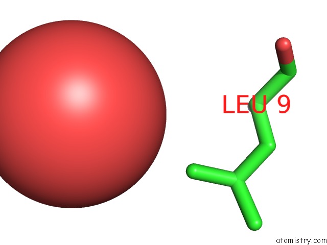
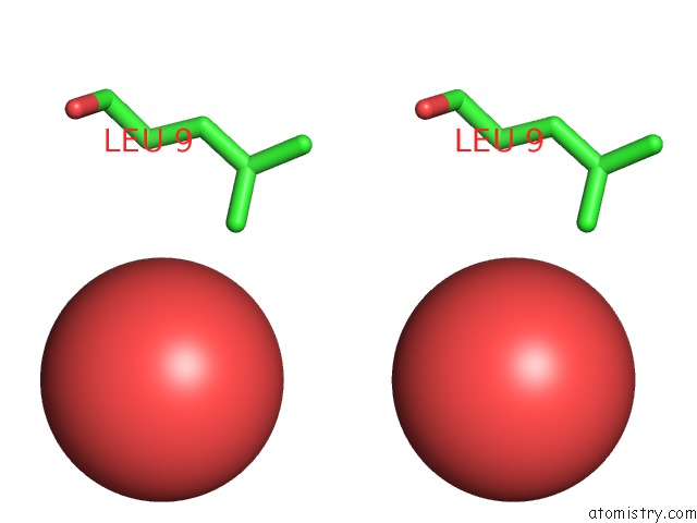
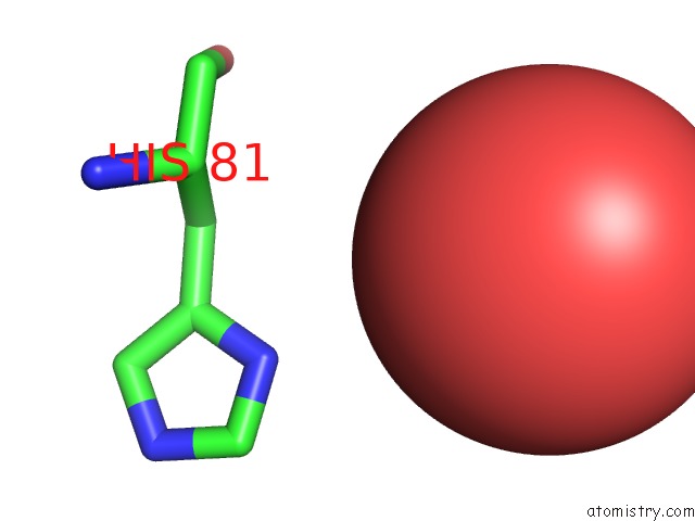
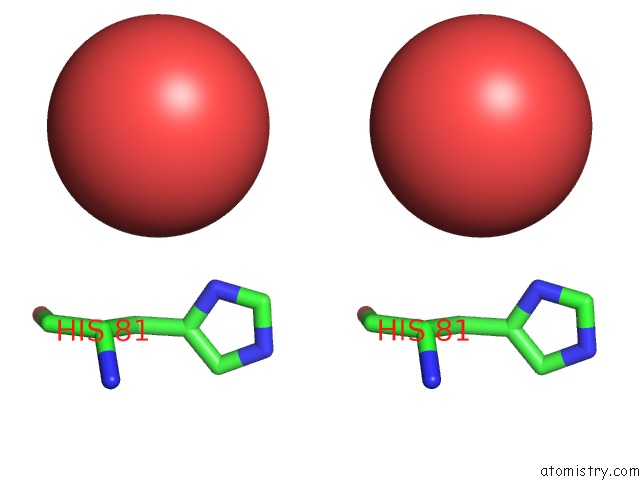
Ruthenium binding site 1 out of 6 in 3ase
Go back to
Ruthenium binding site 1 out
of 6 in the Crystal Structure of Zinc Myoglobin Soaked with RU3O Cluster
Mono view
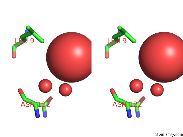
Stereo pair view
Mono view
Stereo pair view
A full contact list of Ruthenium with other atoms in the Ru binding
site number 1 of Crystal Structure of Zinc Myoglobin Soaked with RU3O Cluster within 5.0Å range:
|
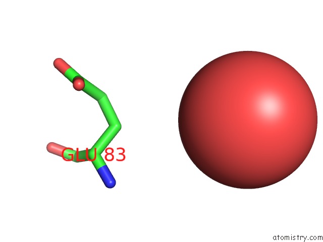
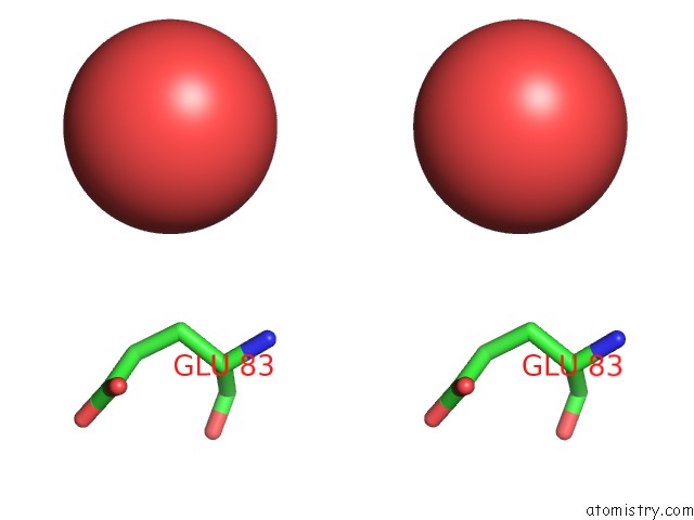
Ruthenium binding site 2 out of 6 in 3ase
Go back to
Ruthenium binding site 2 out
of 6 in the Crystal Structure of Zinc Myoglobin Soaked with RU3O Cluster
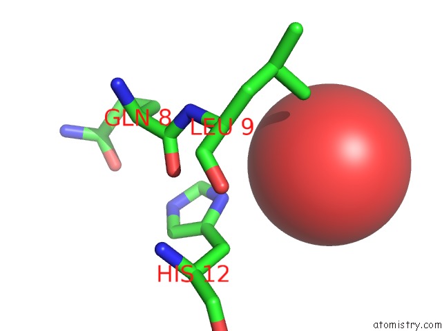
Mono view
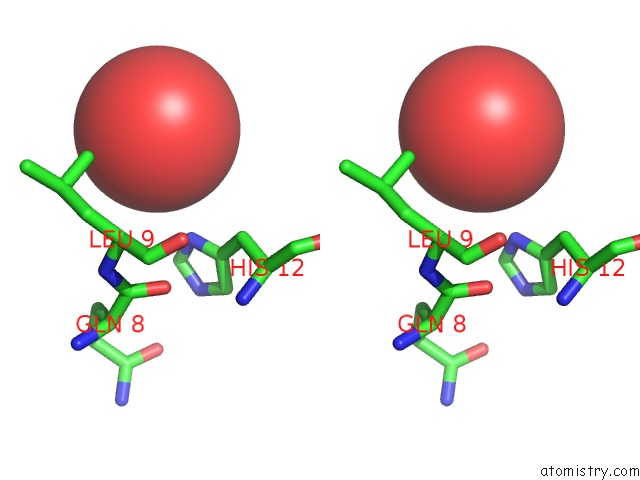
Stereo pair view
Mono view
Stereo pair view
A full contact list of Ruthenium with other atoms in the Ru binding
site number 2 of Crystal Structure of Zinc Myoglobin Soaked with RU3O Cluster within 5.0Å range:
|
Ruthenium binding site 3 out of 6 in 3ase
Go back to
Ruthenium binding site 3 out
of 6 in the Crystal Structure of Zinc Myoglobin Soaked with RU3O Cluster
Mono view
Stereo pair view
Mono view
Stereo pair view
A full contact list of Ruthenium with other atoms in the Ru binding
site number 3 of Crystal Structure of Zinc Myoglobin Soaked with RU3O Cluster within 5.0Å range:
|
Ruthenium binding site 4 out of 6 in 3ase
Go back to
Ruthenium binding site 4 out
of 6 in the Crystal Structure of Zinc Myoglobin Soaked with RU3O Cluster
Mono view
Stereo pair view
Mono view
Stereo pair view
A full contact list of Ruthenium with other atoms in the Ru binding
site number 4 of Crystal Structure of Zinc Myoglobin Soaked with RU3O Cluster within 5.0Å range:
|
Ruthenium binding site 5 out of 6 in 3ase
Go back to
Ruthenium binding site 5 out
of 6 in the Crystal Structure of Zinc Myoglobin Soaked with RU3O Cluster
Mono view
Stereo pair view
Mono view
Stereo pair view
A full contact list of Ruthenium with other atoms in the Ru binding
site number 5 of Crystal Structure of Zinc Myoglobin Soaked with RU3O Cluster within 5.0Å range:
|
Ruthenium binding site 6 out of 6 in 3ase
Go back to
Ruthenium binding site 6 out
of 6 in the Crystal Structure of Zinc Myoglobin Soaked with RU3O Cluster
Mono view
Stereo pair view
Mono view
Stereo pair view
A full contact list of Ruthenium with other atoms in the Ru binding
site number 6 of Crystal Structure of Zinc Myoglobin Soaked with RU3O Cluster within 5.0Å range:
|
Reference:
T.Koshiyama,
M.Shirai,
T.Hikage,
H.Tabe,
K.Tanaka,
S.Kitagawa,
T.Ueno.
Post-Crystal Engineering of Zinc-Substituted Myoglobin to Construct A Long-Lived Photoinduced Charge-Separation System Angew.Chem.Int.Ed.Engl. 2011.
ISSN: ESSN 1521-3773
PubMed: 21495132
DOI: 10.1002/ANIE.201008004
Page generated: Tue Aug 19 00:50:10 2025
ISSN: ESSN 1521-3773
PubMed: 21495132
DOI: 10.1002/ANIE.201008004
Last articles
Mn in 9LJUMn in 9LJW
Mn in 9LJS
Mn in 9LJR
Mn in 9LJT
Mn in 9LJV
Mg in 9UA2
Mg in 9R96
Mg in 9VM1
Mg in 9P01