Ruthenium »
PDB 8p6b-8s90 »
8ph6 »
Ruthenium in PDB 8ph6: X-Ray Structure of the Adduct Formed Upon Reaction of Lysozyme with K2[RU2(Dphf)(CO3)3] in Condition B
Enzymatic activity of X-Ray Structure of the Adduct Formed Upon Reaction of Lysozyme with K2[RU2(Dphf)(CO3)3] in Condition B
All present enzymatic activity of X-Ray Structure of the Adduct Formed Upon Reaction of Lysozyme with K2[RU2(Dphf)(CO3)3] in Condition B:
3.2.1.17;
3.2.1.17;
Protein crystallography data
The structure of X-Ray Structure of the Adduct Formed Upon Reaction of Lysozyme with K2[RU2(Dphf)(CO3)3] in Condition B, PDB code: 8ph6
was solved by
A.Teran,
G.Ferraro,
A.Merlino,
with X-Ray Crystallography technique. A brief refinement statistics is given in the table below:
| Resolution Low / High (Å) | 54.36 / 1.07 |
| Space group | P 43 21 2 |
| Cell size a, b, c (Å), α, β, γ (°) | 76.88, 76.88, 38.61, 90, 90, 90 |
| R / Rfree (%) | 17 / 19.3 |
Ruthenium Binding Sites:
The binding sites of Ruthenium atom in the X-Ray Structure of the Adduct Formed Upon Reaction of Lysozyme with K2[RU2(Dphf)(CO3)3] in Condition B
(pdb code 8ph6). This binding sites where shown within
5.0 Angstroms radius around Ruthenium atom.
In total 10 binding sites of Ruthenium where determined in the X-Ray Structure of the Adduct Formed Upon Reaction of Lysozyme with K2[RU2(Dphf)(CO3)3] in Condition B, PDB code: 8ph6:
Jump to Ruthenium binding site number: 1; 2; 3; 4; 5; 6; 7; 8; 9; 10;
In total 10 binding sites of Ruthenium where determined in the X-Ray Structure of the Adduct Formed Upon Reaction of Lysozyme with K2[RU2(Dphf)(CO3)3] in Condition B, PDB code: 8ph6:
Jump to Ruthenium binding site number: 1; 2; 3; 4; 5; 6; 7; 8; 9; 10;
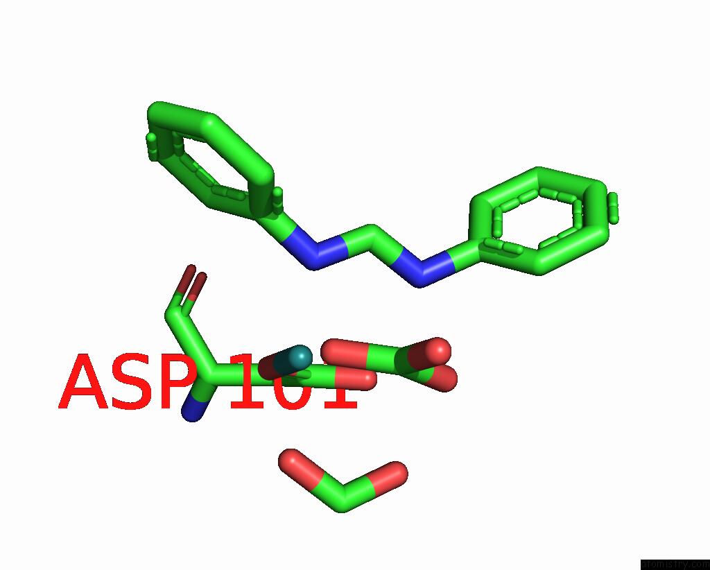
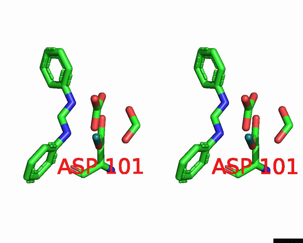
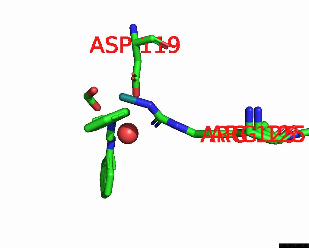
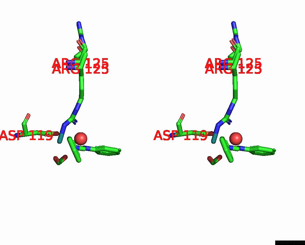
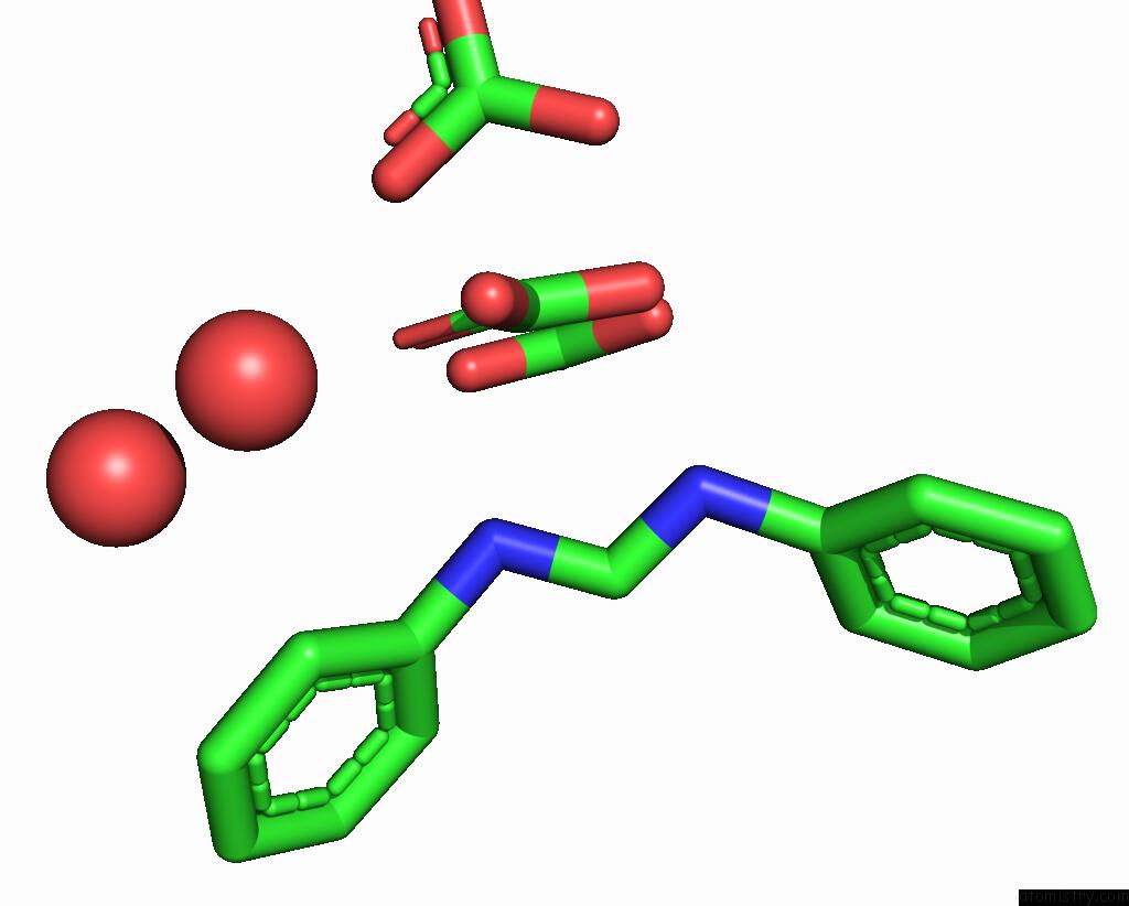
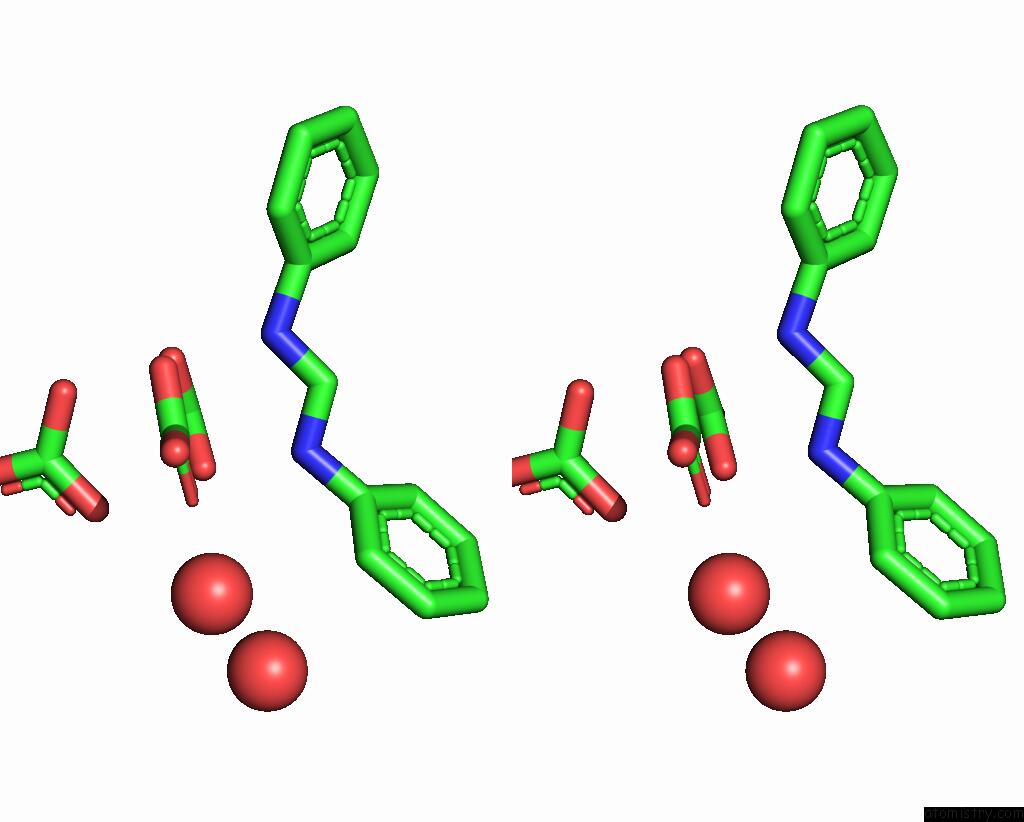
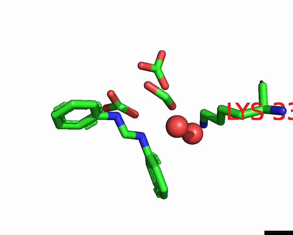
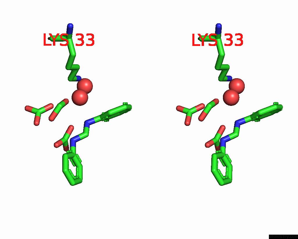
Ruthenium binding site 1 out of 10 in 8ph6
Go back to
Ruthenium binding site 1 out
of 10 in the X-Ray Structure of the Adduct Formed Upon Reaction of Lysozyme with K2[RU2(Dphf)(CO3)3] in Condition B
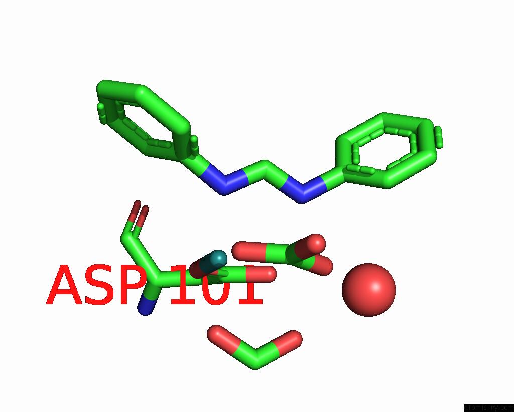
Mono view
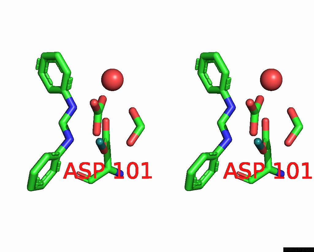
Stereo pair view
Mono view
Stereo pair view
A full contact list of Ruthenium with other atoms in the Ru binding
site number 1 of X-Ray Structure of the Adduct Formed Upon Reaction of Lysozyme with K2[RU2(Dphf)(CO3)3] in Condition B within 5.0Å range:
|
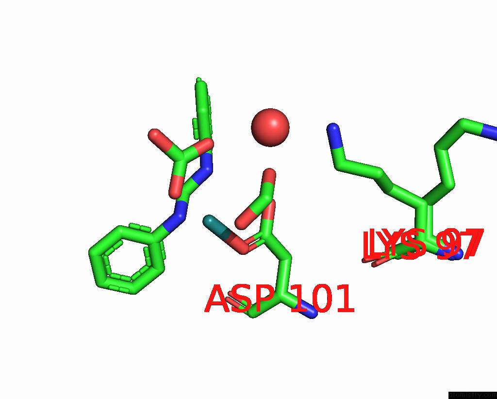
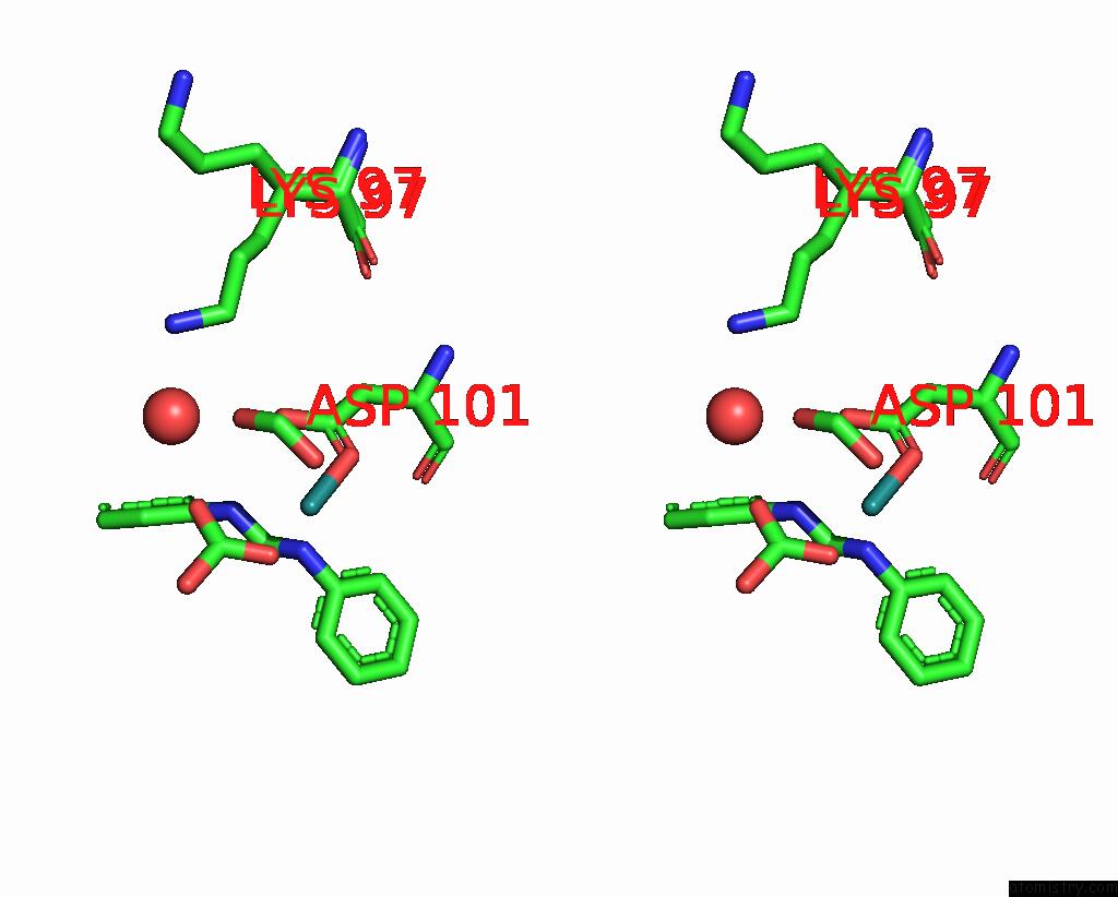
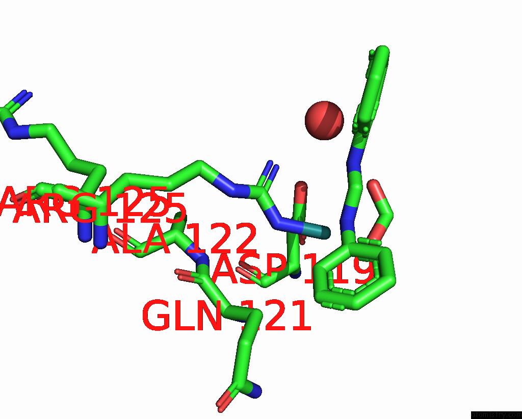
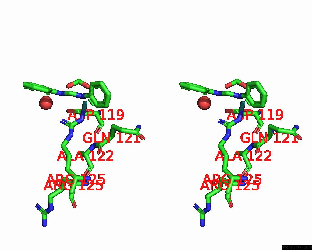
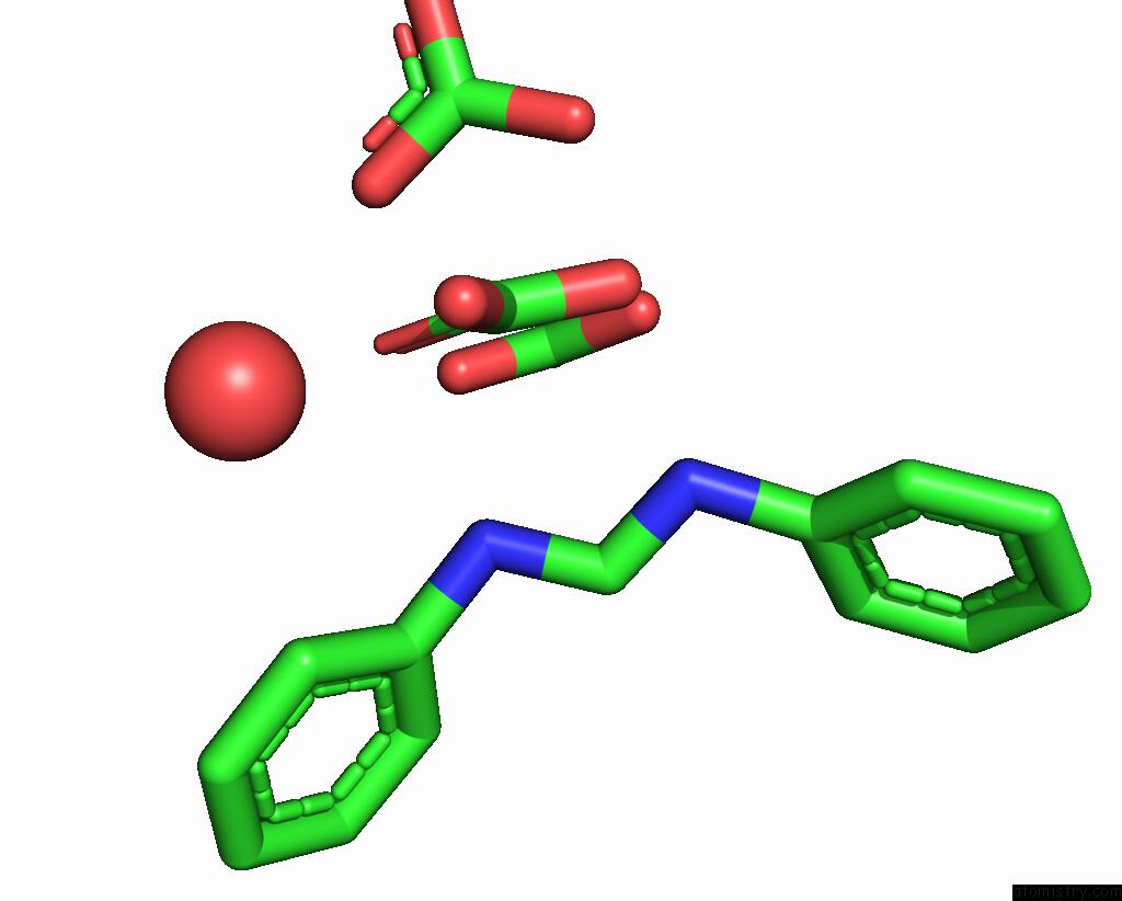
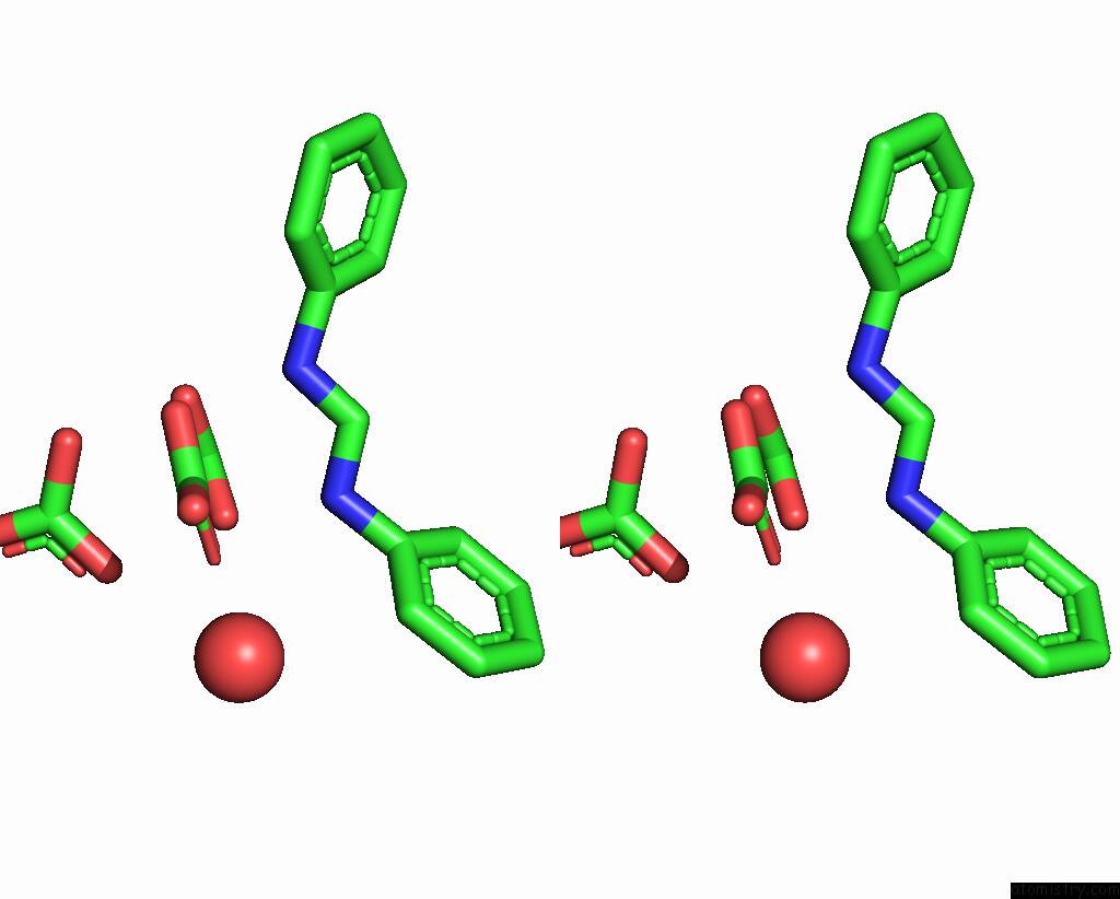
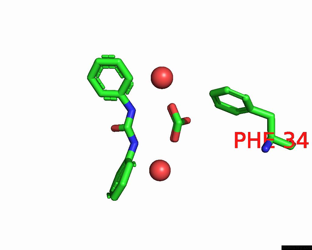
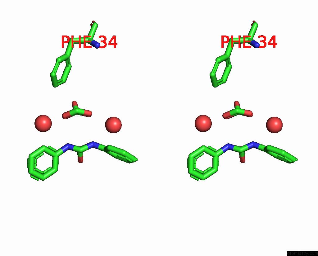
Ruthenium binding site 2 out of 10 in 8ph6
Go back to
Ruthenium binding site 2 out
of 10 in the X-Ray Structure of the Adduct Formed Upon Reaction of Lysozyme with K2[RU2(Dphf)(CO3)3] in Condition B

Mono view

Stereo pair view
Mono view
Stereo pair view
A full contact list of Ruthenium with other atoms in the Ru binding
site number 2 of X-Ray Structure of the Adduct Formed Upon Reaction of Lysozyme with K2[RU2(Dphf)(CO3)3] in Condition B within 5.0Å range:
|
Ruthenium binding site 3 out of 10 in 8ph6
Go back to
Ruthenium binding site 3 out
of 10 in the X-Ray Structure of the Adduct Formed Upon Reaction of Lysozyme with K2[RU2(Dphf)(CO3)3] in Condition B
Mono view
Stereo pair view
Mono view
Stereo pair view
A full contact list of Ruthenium with other atoms in the Ru binding
site number 3 of X-Ray Structure of the Adduct Formed Upon Reaction of Lysozyme with K2[RU2(Dphf)(CO3)3] in Condition B within 5.0Å range:
|
Ruthenium binding site 4 out of 10 in 8ph6
Go back to
Ruthenium binding site 4 out
of 10 in the X-Ray Structure of the Adduct Formed Upon Reaction of Lysozyme with K2[RU2(Dphf)(CO3)3] in Condition B
Mono view
Stereo pair view
Mono view
Stereo pair view
A full contact list of Ruthenium with other atoms in the Ru binding
site number 4 of X-Ray Structure of the Adduct Formed Upon Reaction of Lysozyme with K2[RU2(Dphf)(CO3)3] in Condition B within 5.0Å range:
|
Ruthenium binding site 5 out of 10 in 8ph6
Go back to
Ruthenium binding site 5 out
of 10 in the X-Ray Structure of the Adduct Formed Upon Reaction of Lysozyme with K2[RU2(Dphf)(CO3)3] in Condition B
Mono view
Stereo pair view
Mono view
Stereo pair view
A full contact list of Ruthenium with other atoms in the Ru binding
site number 5 of X-Ray Structure of the Adduct Formed Upon Reaction of Lysozyme with K2[RU2(Dphf)(CO3)3] in Condition B within 5.0Å range:
|
Ruthenium binding site 6 out of 10 in 8ph6
Go back to
Ruthenium binding site 6 out
of 10 in the X-Ray Structure of the Adduct Formed Upon Reaction of Lysozyme with K2[RU2(Dphf)(CO3)3] in Condition B
Mono view
Stereo pair view
Mono view
Stereo pair view
A full contact list of Ruthenium with other atoms in the Ru binding
site number 6 of X-Ray Structure of the Adduct Formed Upon Reaction of Lysozyme with K2[RU2(Dphf)(CO3)3] in Condition B within 5.0Å range:
|
Ruthenium binding site 7 out of 10 in 8ph6
Go back to
Ruthenium binding site 7 out
of 10 in the X-Ray Structure of the Adduct Formed Upon Reaction of Lysozyme with K2[RU2(Dphf)(CO3)3] in Condition B
Mono view
Stereo pair view
Mono view
Stereo pair view
A full contact list of Ruthenium with other atoms in the Ru binding
site number 7 of X-Ray Structure of the Adduct Formed Upon Reaction of Lysozyme with K2[RU2(Dphf)(CO3)3] in Condition B within 5.0Å range:
|
Ruthenium binding site 8 out of 10 in 8ph6
Go back to
Ruthenium binding site 8 out
of 10 in the X-Ray Structure of the Adduct Formed Upon Reaction of Lysozyme with K2[RU2(Dphf)(CO3)3] in Condition B
Mono view
Stereo pair view
Mono view
Stereo pair view
A full contact list of Ruthenium with other atoms in the Ru binding
site number 8 of X-Ray Structure of the Adduct Formed Upon Reaction of Lysozyme with K2[RU2(Dphf)(CO3)3] in Condition B within 5.0Å range:
|
Ruthenium binding site 9 out of 10 in 8ph6
Go back to
Ruthenium binding site 9 out
of 10 in the X-Ray Structure of the Adduct Formed Upon Reaction of Lysozyme with K2[RU2(Dphf)(CO3)3] in Condition B
Mono view
Stereo pair view
Mono view
Stereo pair view
A full contact list of Ruthenium with other atoms in the Ru binding
site number 9 of X-Ray Structure of the Adduct Formed Upon Reaction of Lysozyme with K2[RU2(Dphf)(CO3)3] in Condition B within 5.0Å range:
|
Ruthenium binding site 10 out of 10 in 8ph6
Go back to
Ruthenium binding site 10 out
of 10 in the X-Ray Structure of the Adduct Formed Upon Reaction of Lysozyme with K2[RU2(Dphf)(CO3)3] in Condition B
Mono view
Stereo pair view
Mono view
Stereo pair view
A full contact list of Ruthenium with other atoms in the Ru binding
site number 10 of X-Ray Structure of the Adduct Formed Upon Reaction of Lysozyme with K2[RU2(Dphf)(CO3)3] in Condition B within 5.0Å range:
|
Reference:
A.Teran,
G.Ferraro,
P.Imbimbo,
A.E.Sanchez-Pelaez,
D.M.Monti,
S.Herrero,
A.Merlino.
Steric Hindrance and Charge Influence on the Cytotoxic Activity and Protein Binding Properties of Diruthenium Complexes. Int.J.Biol.Macromol. 26666 2023.
ISSN: ISSN 0141-8130
PubMed: 37660867
DOI: 10.1016/J.IJBIOMAC.2023.126666
Page generated: Thu Oct 10 13:14:15 2024
ISSN: ISSN 0141-8130
PubMed: 37660867
DOI: 10.1016/J.IJBIOMAC.2023.126666
Last articles
Cl in 2Y2MCl in 2Y2G
Cl in 2Y2K
Cl in 2Y2H
Cl in 2Y2J
Cl in 2Y2I
Cl in 2Y1X
Cl in 2Y1G
Cl in 2Y1F
Cl in 2Y05