Ruthenium »
PDB 8p6b-8s90 »
8pfu »
Ruthenium in PDB 8pfu: X-Ray Structure of the Adduct Formed Upon Reaction of Lysozyme with K3[RU2(CO3)4] in Condition A
Enzymatic activity of X-Ray Structure of the Adduct Formed Upon Reaction of Lysozyme with K3[RU2(CO3)4] in Condition A
All present enzymatic activity of X-Ray Structure of the Adduct Formed Upon Reaction of Lysozyme with K3[RU2(CO3)4] in Condition A:
3.2.1.17;
3.2.1.17;
Protein crystallography data
The structure of X-Ray Structure of the Adduct Formed Upon Reaction of Lysozyme with K3[RU2(CO3)4] in Condition A, PDB code: 8pfu
was solved by
A.Teran,
G.Ferraro,
A.Merlino,
with X-Ray Crystallography technique. A brief refinement statistics is given in the table below:
| Resolution Low / High (Å) | 55.20 / 1.18 |
| Space group | P 43 21 2 |
| Cell size a, b, c (Å), α, β, γ (°) | 78.06, 78.06, 37.47, 90, 90, 90 |
| R / Rfree (%) | 17.9 / 20.2 |
Other elements in 8pfu:
The structure of X-Ray Structure of the Adduct Formed Upon Reaction of Lysozyme with K3[RU2(CO3)4] in Condition A also contains other interesting chemical elements:
| Sodium | (Na) | 1 atom |
Ruthenium Binding Sites:
The binding sites of Ruthenium atom in the X-Ray Structure of the Adduct Formed Upon Reaction of Lysozyme with K3[RU2(CO3)4] in Condition A
(pdb code 8pfu). This binding sites where shown within
5.0 Angstroms radius around Ruthenium atom.
In total 8 binding sites of Ruthenium where determined in the X-Ray Structure of the Adduct Formed Upon Reaction of Lysozyme with K3[RU2(CO3)4] in Condition A, PDB code: 8pfu:
Jump to Ruthenium binding site number: 1; 2; 3; 4; 5; 6; 7; 8;
In total 8 binding sites of Ruthenium where determined in the X-Ray Structure of the Adduct Formed Upon Reaction of Lysozyme with K3[RU2(CO3)4] in Condition A, PDB code: 8pfu:
Jump to Ruthenium binding site number: 1; 2; 3; 4; 5; 6; 7; 8;
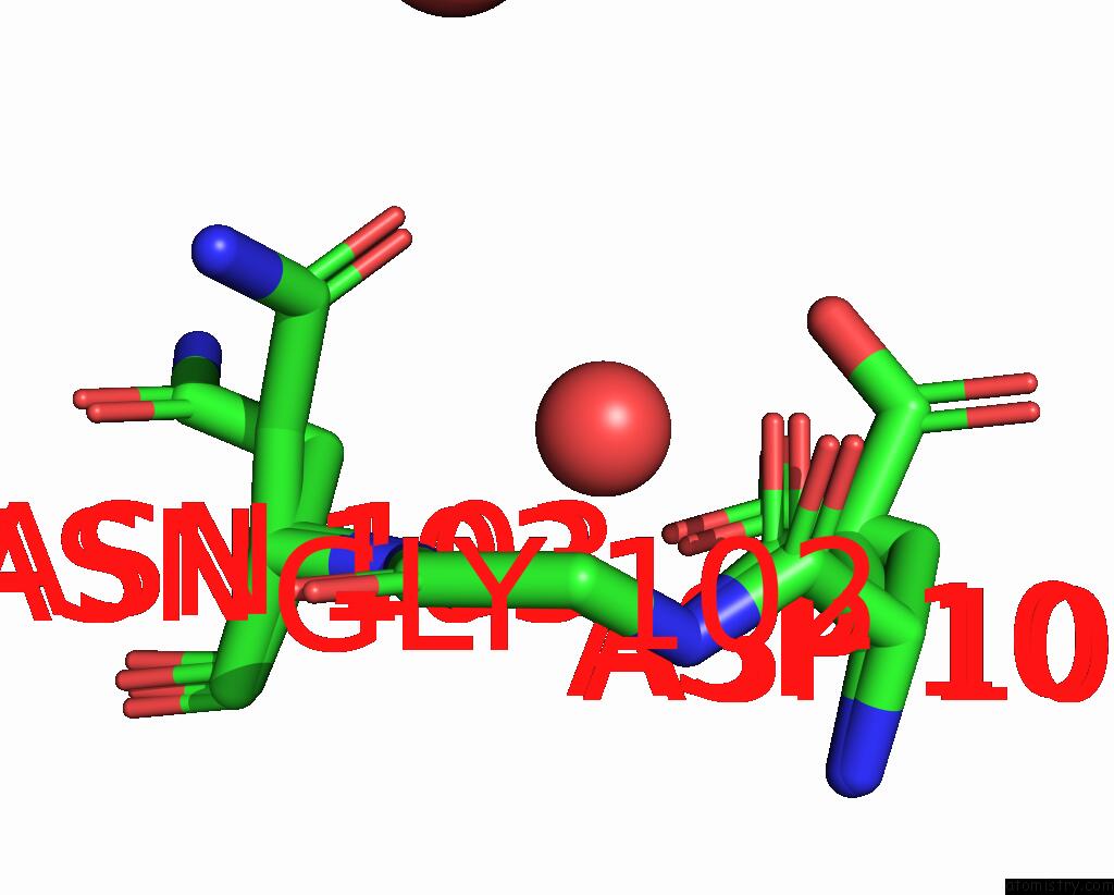
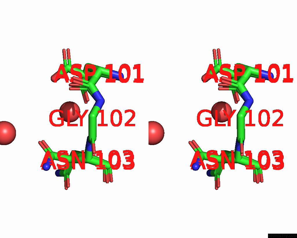
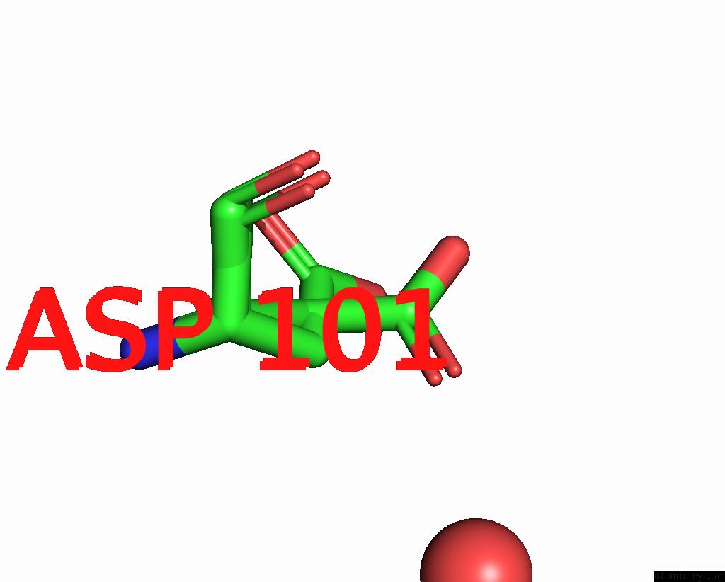
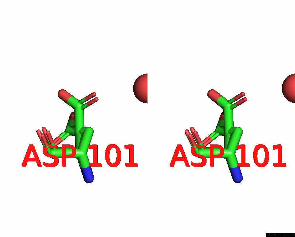
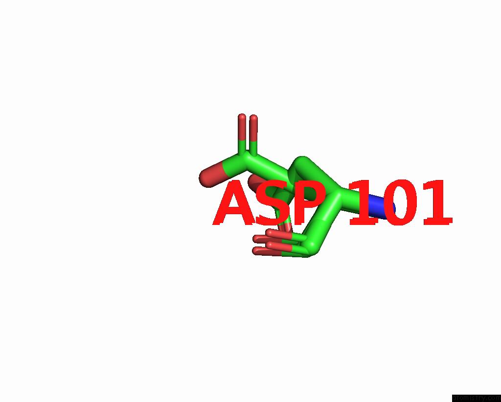
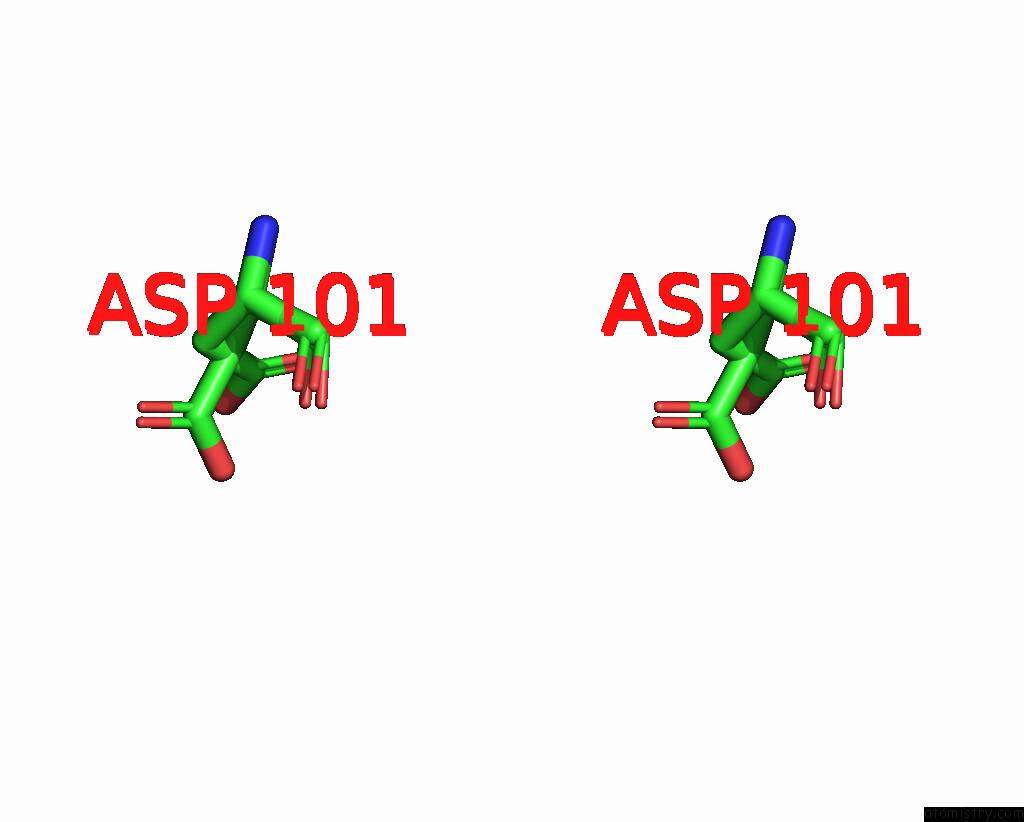
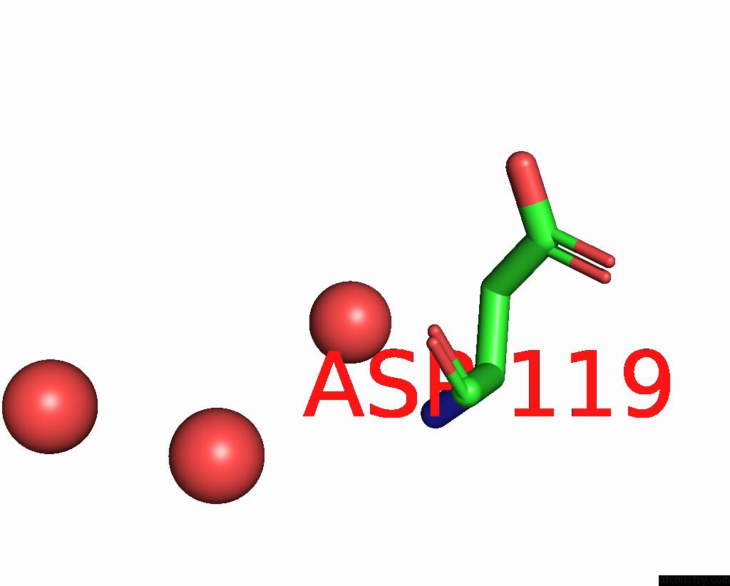
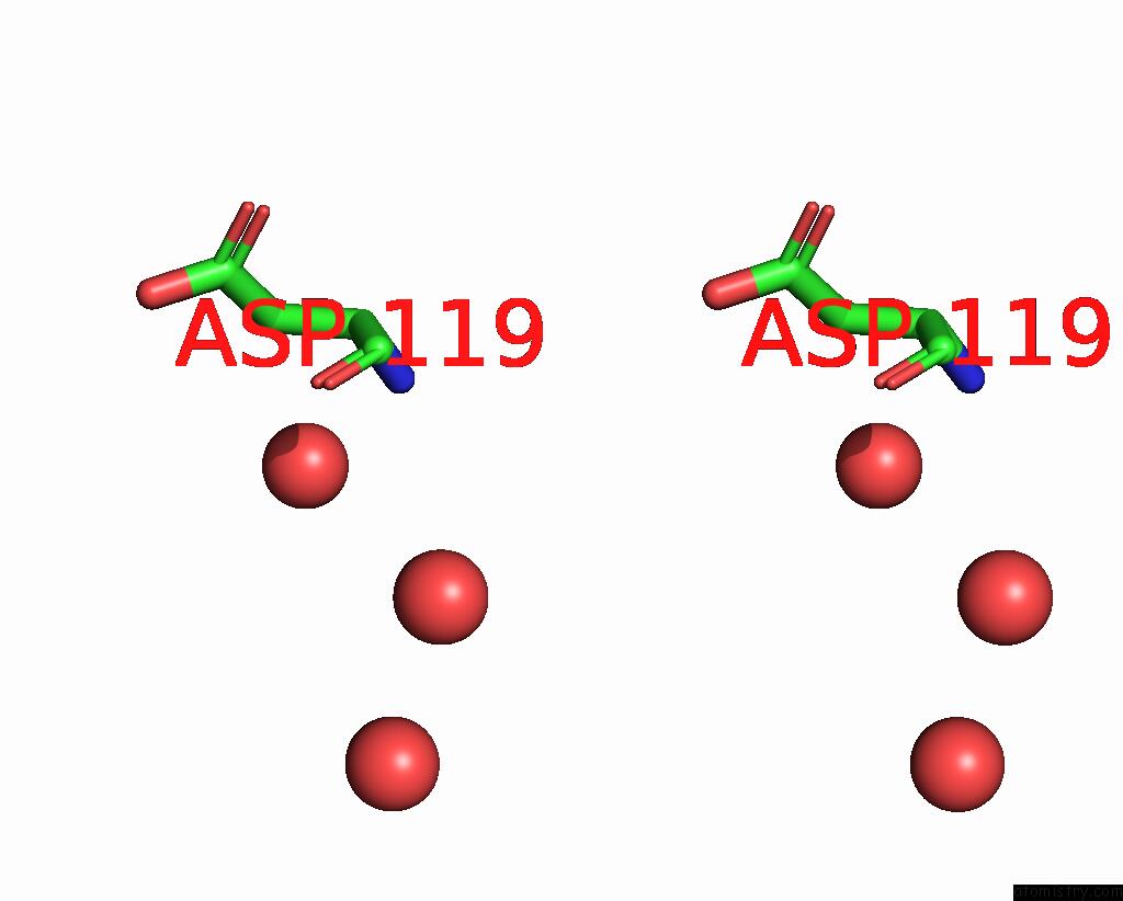
Ruthenium binding site 1 out of 8 in 8pfu
Go back to
Ruthenium binding site 1 out
of 8 in the X-Ray Structure of the Adduct Formed Upon Reaction of Lysozyme with K3[RU2(CO3)4] in Condition A
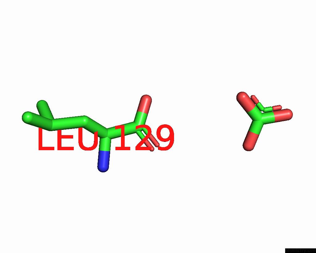
Mono view
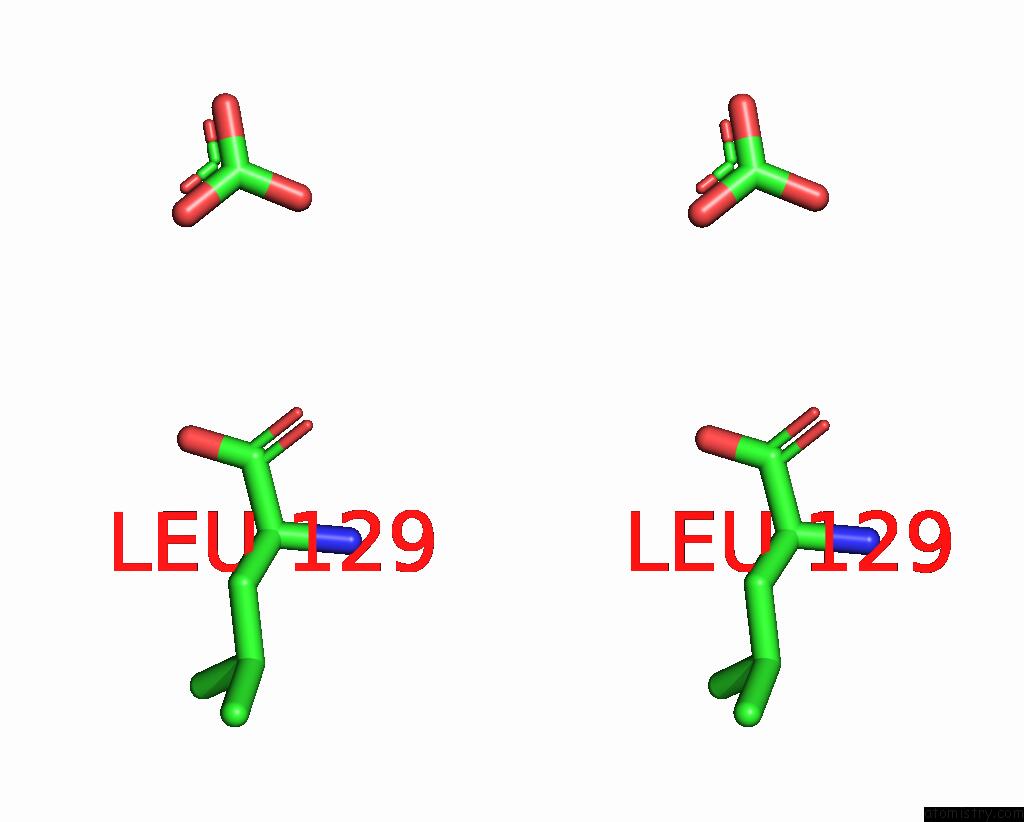
Stereo pair view
Mono view
Stereo pair view
A full contact list of Ruthenium with other atoms in the Ru binding
site number 1 of X-Ray Structure of the Adduct Formed Upon Reaction of Lysozyme with K3[RU2(CO3)4] in Condition A within 5.0Å range:
|
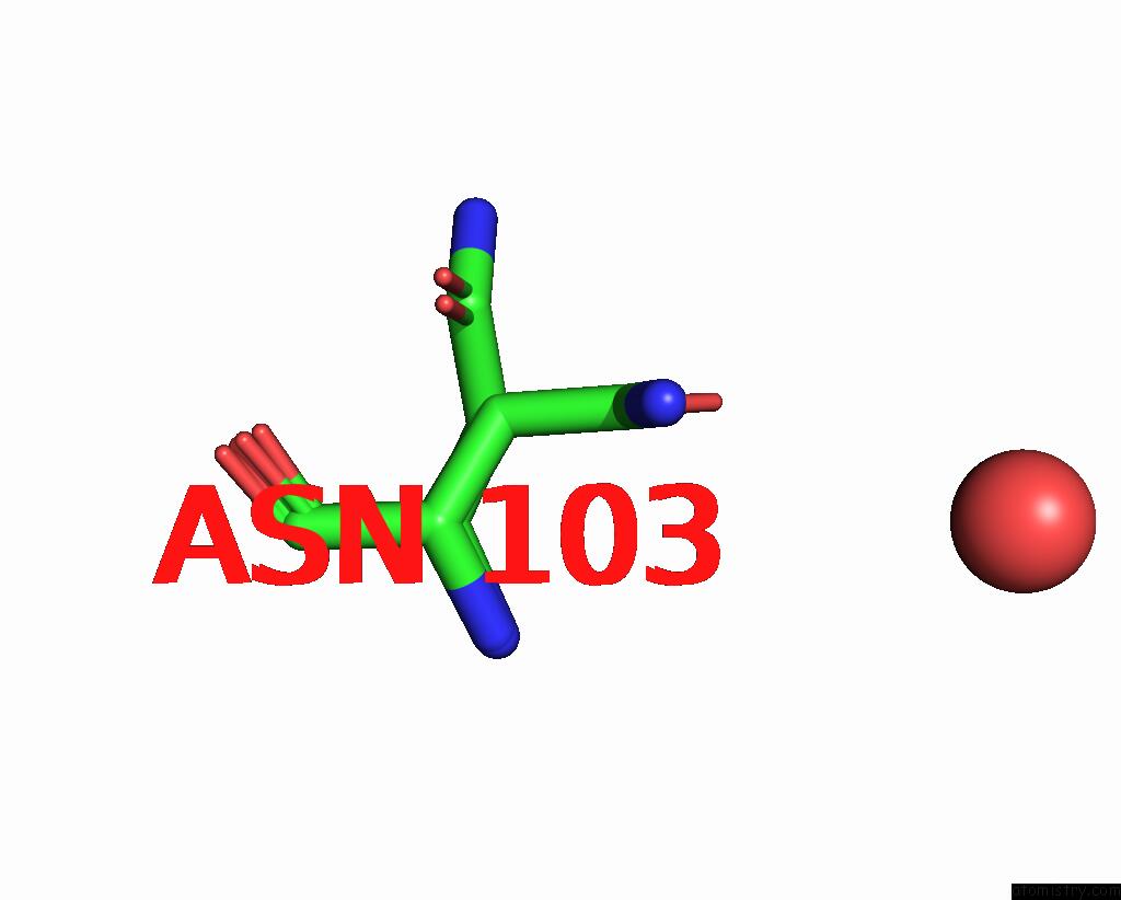
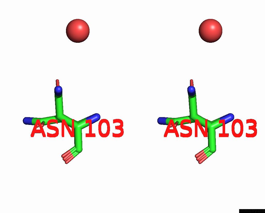
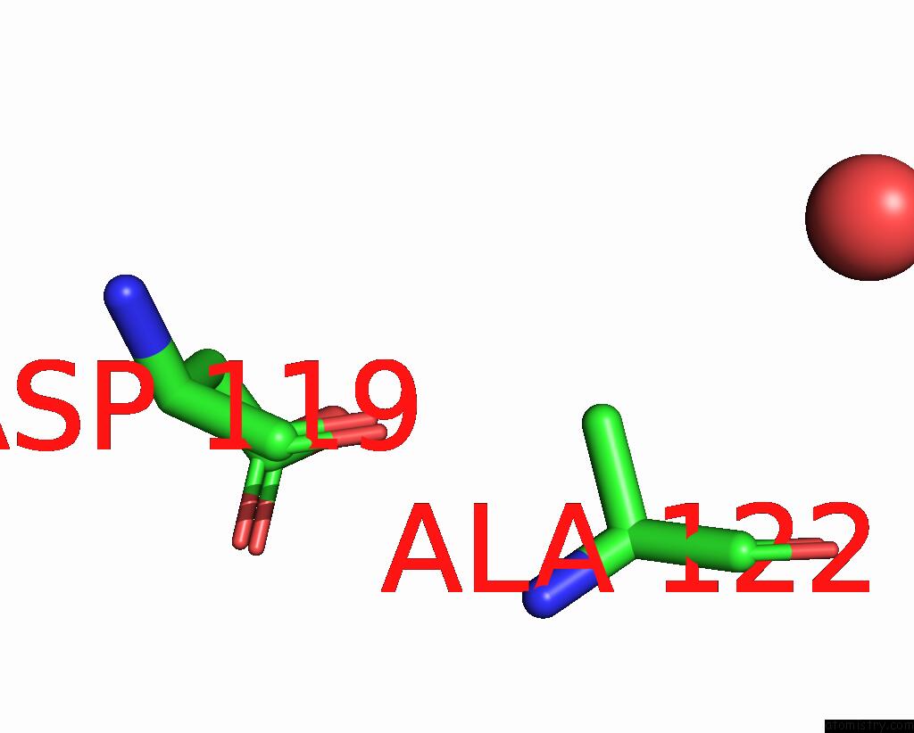
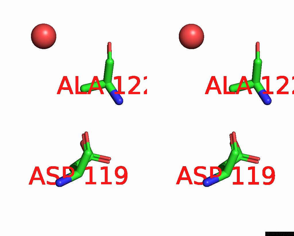
Ruthenium binding site 2 out of 8 in 8pfu
Go back to
Ruthenium binding site 2 out
of 8 in the X-Ray Structure of the Adduct Formed Upon Reaction of Lysozyme with K3[RU2(CO3)4] in Condition A
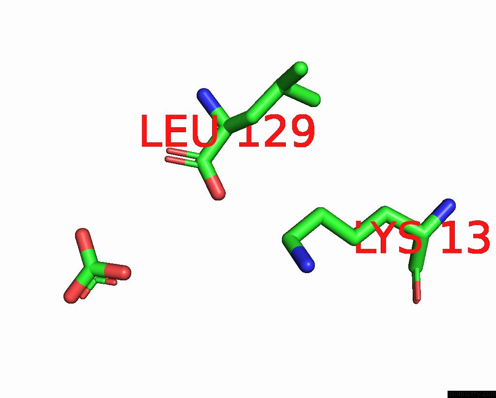
Mono view
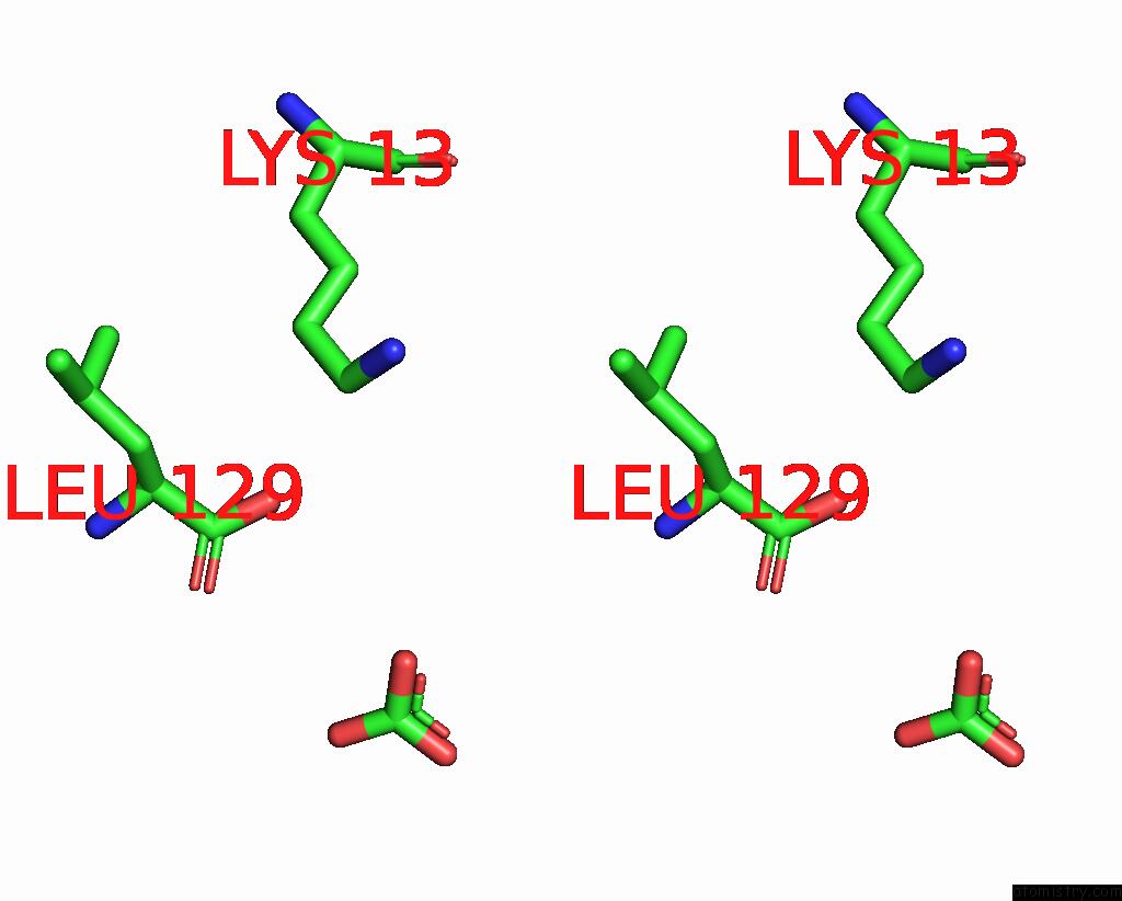
Stereo pair view
Mono view
Stereo pair view
A full contact list of Ruthenium with other atoms in the Ru binding
site number 2 of X-Ray Structure of the Adduct Formed Upon Reaction of Lysozyme with K3[RU2(CO3)4] in Condition A within 5.0Å range:
|
Ruthenium binding site 3 out of 8 in 8pfu
Go back to
Ruthenium binding site 3 out
of 8 in the X-Ray Structure of the Adduct Formed Upon Reaction of Lysozyme with K3[RU2(CO3)4] in Condition A
Mono view
Stereo pair view
Mono view
Stereo pair view
A full contact list of Ruthenium with other atoms in the Ru binding
site number 3 of X-Ray Structure of the Adduct Formed Upon Reaction of Lysozyme with K3[RU2(CO3)4] in Condition A within 5.0Å range:
|
Ruthenium binding site 4 out of 8 in 8pfu
Go back to
Ruthenium binding site 4 out
of 8 in the X-Ray Structure of the Adduct Formed Upon Reaction of Lysozyme with K3[RU2(CO3)4] in Condition A
Mono view
Stereo pair view
Mono view
Stereo pair view
A full contact list of Ruthenium with other atoms in the Ru binding
site number 4 of X-Ray Structure of the Adduct Formed Upon Reaction of Lysozyme with K3[RU2(CO3)4] in Condition A within 5.0Å range:
|
Ruthenium binding site 5 out of 8 in 8pfu
Go back to
Ruthenium binding site 5 out
of 8 in the X-Ray Structure of the Adduct Formed Upon Reaction of Lysozyme with K3[RU2(CO3)4] in Condition A
Mono view
Stereo pair view
Mono view
Stereo pair view
A full contact list of Ruthenium with other atoms in the Ru binding
site number 5 of X-Ray Structure of the Adduct Formed Upon Reaction of Lysozyme with K3[RU2(CO3)4] in Condition A within 5.0Å range:
|
Ruthenium binding site 6 out of 8 in 8pfu
Go back to
Ruthenium binding site 6 out
of 8 in the X-Ray Structure of the Adduct Formed Upon Reaction of Lysozyme with K3[RU2(CO3)4] in Condition A
Mono view
Stereo pair view
Mono view
Stereo pair view
A full contact list of Ruthenium with other atoms in the Ru binding
site number 6 of X-Ray Structure of the Adduct Formed Upon Reaction of Lysozyme with K3[RU2(CO3)4] in Condition A within 5.0Å range:
|
Ruthenium binding site 7 out of 8 in 8pfu
Go back to
Ruthenium binding site 7 out
of 8 in the X-Ray Structure of the Adduct Formed Upon Reaction of Lysozyme with K3[RU2(CO3)4] in Condition A
Mono view
Stereo pair view
Mono view
Stereo pair view
A full contact list of Ruthenium with other atoms in the Ru binding
site number 7 of X-Ray Structure of the Adduct Formed Upon Reaction of Lysozyme with K3[RU2(CO3)4] in Condition A within 5.0Å range:
|
Ruthenium binding site 8 out of 8 in 8pfu
Go back to
Ruthenium binding site 8 out
of 8 in the X-Ray Structure of the Adduct Formed Upon Reaction of Lysozyme with K3[RU2(CO3)4] in Condition A
Mono view
Stereo pair view
Mono view
Stereo pair view
A full contact list of Ruthenium with other atoms in the Ru binding
site number 8 of X-Ray Structure of the Adduct Formed Upon Reaction of Lysozyme with K3[RU2(CO3)4] in Condition A within 5.0Å range:
|
Reference:
S.Herrero Dominguez,
A.Teran,
G.Ferraro,
A.E.Sanchez-Pelaez,
A.Merlino.
Charge Effect in Protein Metalation Reactions By Diruthenium Complexes Inorg Chem Front 2023.
DOI: 10.1039/D3QI01192E
Page generated: Thu Oct 10 13:14:08 2024
DOI: 10.1039/D3QI01192E
Last articles
Zn in 9MJ5Zn in 9HNW
Zn in 9G0L
Zn in 9FNE
Zn in 9DZN
Zn in 9E0I
Zn in 9D32
Zn in 9DAK
Zn in 8ZXC
Zn in 8ZUF