Ruthenium »
PDB 4l55-5ira »
5ifo »
Ruthenium in PDB 5ifo: X-Ray Structure of Hsa-Myr-KP1019
Protein crystallography data
The structure of X-Ray Structure of Hsa-Myr-KP1019, PDB code: 5ifo
was solved by
A.Bijelic,
S.Theiner,
B.K.Keppler,
A.Rompel,
with X-Ray Crystallography technique. A brief refinement statistics is given in the table below:
| Resolution Low / High (Å) | 45.84 / 3.20 |
| Space group | C 1 2 1 |
| Cell size a, b, c (Å), α, β, γ (°) | 181.110, 38.060, 94.950, 90.00, 105.06, 90.00 |
| R / Rfree (%) | 24.4 / 26.2 |
Ruthenium Binding Sites:
The binding sites of Ruthenium atom in the X-Ray Structure of Hsa-Myr-KP1019
(pdb code 5ifo). This binding sites where shown within
5.0 Angstroms radius around Ruthenium atom.
In total 2 binding sites of Ruthenium where determined in the X-Ray Structure of Hsa-Myr-KP1019, PDB code: 5ifo:
Jump to Ruthenium binding site number: 1; 2;
In total 2 binding sites of Ruthenium where determined in the X-Ray Structure of Hsa-Myr-KP1019, PDB code: 5ifo:
Jump to Ruthenium binding site number: 1; 2;
Ruthenium binding site 1 out of 2 in 5ifo
Go back to
Ruthenium binding site 1 out
of 2 in the X-Ray Structure of Hsa-Myr-KP1019
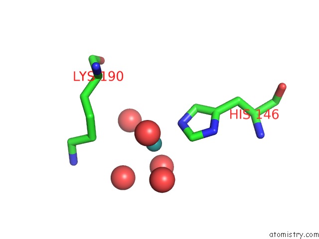
Mono view
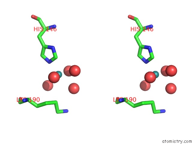
Stereo pair view
Mono view
Stereo pair view
A full contact list of Ruthenium with other atoms in the Ru binding
site number 1 of X-Ray Structure of Hsa-Myr-KP1019 within 5.0Å range:
|
Ruthenium binding site 2 out of 2 in 5ifo
Go back to
Ruthenium binding site 2 out
of 2 in the X-Ray Structure of Hsa-Myr-KP1019
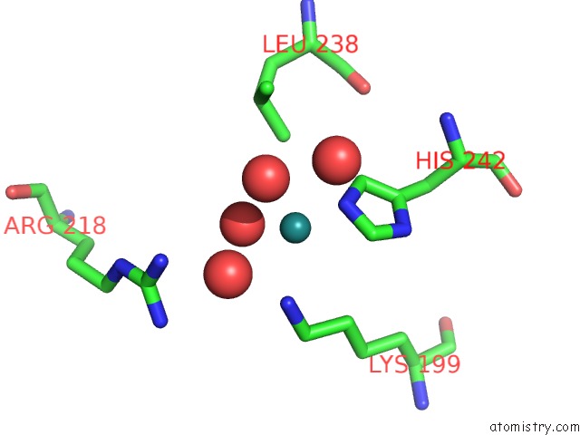
Mono view
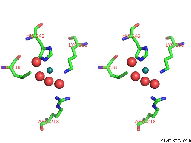
Stereo pair view
Mono view
Stereo pair view
A full contact list of Ruthenium with other atoms in the Ru binding
site number 2 of X-Ray Structure of Hsa-Myr-KP1019 within 5.0Å range:
|
Reference:
A.Bijelic,
S.Theiner,
B.K.Keppler,
A.Rompel.
X-Ray Structure Analysis of Indazolium Trans-[Tetrachlorobis(1H-Indazole)Ruthenate(III)] (KP1019) Bound to Human Serum Albumin Reveals Two Ruthenium Binding Sites and Provides Insights Into the Drug Binding Mechanism. J.Med.Chem. V. 59 5894 2016.
ISSN: ISSN 0022-2623
PubMed: 27196130
DOI: 10.1021/ACS.JMEDCHEM.6B00600
Page generated: Thu Oct 10 12:58:53 2024
ISSN: ISSN 0022-2623
PubMed: 27196130
DOI: 10.1021/ACS.JMEDCHEM.6B00600
Last articles
Zn in 9J0NZn in 9J0O
Zn in 9J0P
Zn in 9FJX
Zn in 9EKB
Zn in 9C0F
Zn in 9CAH
Zn in 9CH0
Zn in 9CH3
Zn in 9CH1