Ruthenium »
PDB 4l55-5ira »
5et2 »
Ruthenium in PDB 5et2: Lambda-Ru(Tap)2(Dppz)]2+ Bound to D(Ttggcgccaa)
Protein crystallography data
The structure of Lambda-Ru(Tap)2(Dppz)]2+ Bound to D(Ttggcgccaa), PDB code: 5et2
was solved by
J.P.Hall,
C.J.Cardin,
with X-Ray Crystallography technique. A brief refinement statistics is given in the table below:
| Resolution Low / High (Å) | 23.83 / 1.39 |
| Space group | P 43 21 2 |
| Cell size a, b, c (Å), α, β, γ (°) | 42.380, 42.380, 39.300, 90.00, 90.00, 90.00 |
| R / Rfree (%) | 16.6 / 18.5 |
Other elements in 5et2:
The structure of Lambda-Ru(Tap)2(Dppz)]2+ Bound to D(Ttggcgccaa) also contains other interesting chemical elements:
| Barium | (Ba) | 1 atom |
Ruthenium Binding Sites:
The binding sites of Ruthenium atom in the Lambda-Ru(Tap)2(Dppz)]2+ Bound to D(Ttggcgccaa)
(pdb code 5et2). This binding sites where shown within
5.0 Angstroms radius around Ruthenium atom.
In total only one binding site of Ruthenium was determined in the Lambda-Ru(Tap)2(Dppz)]2+ Bound to D(Ttggcgccaa), PDB code: 5et2:
In total only one binding site of Ruthenium was determined in the Lambda-Ru(Tap)2(Dppz)]2+ Bound to D(Ttggcgccaa), PDB code: 5et2:
Ruthenium binding site 1 out of 1 in 5et2
Go back to
Ruthenium binding site 1 out
of 1 in the Lambda-Ru(Tap)2(Dppz)]2+ Bound to D(Ttggcgccaa)
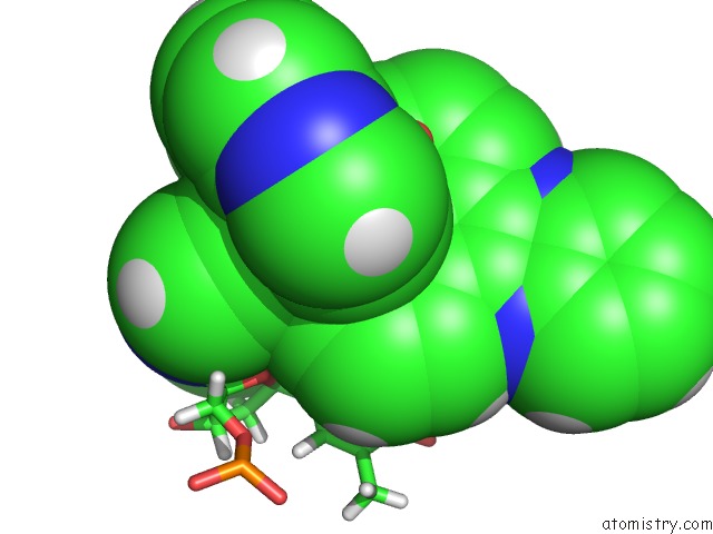
Mono view
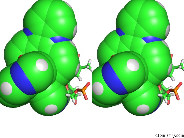
Stereo pair view
Mono view
Stereo pair view
A full contact list of Ruthenium with other atoms in the Ru binding
site number 1 of Lambda-Ru(Tap)2(Dppz)]2+ Bound to D(Ttggcgccaa) within 5.0Å range:
|
Reference:
P.M.Keane,
J.P.Hall,
F.E.Poynton,
B.C.Poulsen,
S.P.Gurung,
I.P.Clark,
I.V.Sazanovich,
M.Towrie,
T.Gunnlaugsson,
S.J.Quinn,
C.J.Cardin,
J.M.Kelly.
Inosine Can Increase Dna'S Susceptibility to Photo-Oxidation By A Ru(II) Complex Due to Structural Change in the Minor Groove. Chemistry V. 23 10344 2017.
ISSN: ISSN 1521-3765
PubMed: 28543779
DOI: 10.1002/CHEM.201701447
Page generated: Thu Oct 10 12:58:27 2024
ISSN: ISSN 1521-3765
PubMed: 28543779
DOI: 10.1002/CHEM.201701447
Last articles
Ca in 2W86Ca in 2W7P
Ca in 2W7O
Ca in 2W67
Ca in 2W68
Ca in 2W4Z
Ca in 2W4Y
Ca in 2W66
Ca in 2W4X
Ca in 2W3O