Ruthenium »
PDB 4l55-5ira »
4x1a »
Ruthenium in PDB 4x1a: Lambda-[Ru(Tap)2(Dppz-10,12-Me)]2+ Bound to D(Tcggcgccga)
Protein crystallography data
The structure of Lambda-[Ru(Tap)2(Dppz-10,12-Me)]2+ Bound to D(Tcggcgccga), PDB code: 4x1a
was solved by
J.P.Hall,
C.J.Cardin,
with X-Ray Crystallography technique. A brief refinement statistics is given in the table below:
| Resolution Low / High (Å) | 17.05 / 0.89 |
| Space group | P 43 21 2 |
| Cell size a, b, c (Å), α, β, γ (°) | 42.250, 42.250, 39.550, 90.00, 90.00, 90.00 |
| R / Rfree (%) | 9.6 / 10.6 |
Other elements in 4x1a:
The structure of Lambda-[Ru(Tap)2(Dppz-10,12-Me)]2+ Bound to D(Tcggcgccga) also contains other interesting chemical elements:
| Barium | (Ba) | 1 atom |
Ruthenium Binding Sites:
The binding sites of Ruthenium atom in the Lambda-[Ru(Tap)2(Dppz-10,12-Me)]2+ Bound to D(Tcggcgccga)
(pdb code 4x1a). This binding sites where shown within
5.0 Angstroms radius around Ruthenium atom.
In total only one binding site of Ruthenium was determined in the Lambda-[Ru(Tap)2(Dppz-10,12-Me)]2+ Bound to D(Tcggcgccga), PDB code: 4x1a:
In total only one binding site of Ruthenium was determined in the Lambda-[Ru(Tap)2(Dppz-10,12-Me)]2+ Bound to D(Tcggcgccga), PDB code: 4x1a:
Ruthenium binding site 1 out of 1 in 4x1a
Go back to
Ruthenium binding site 1 out
of 1 in the Lambda-[Ru(Tap)2(Dppz-10,12-Me)]2+ Bound to D(Tcggcgccga)

Mono view
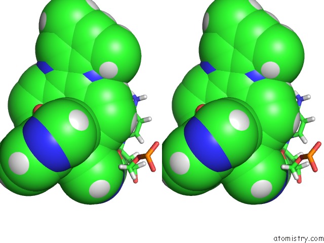
Stereo pair view
Mono view
Stereo pair view
A full contact list of Ruthenium with other atoms in the Ru binding
site number 1 of Lambda-[Ru(Tap)2(Dppz-10,12-Me)]2+ Bound to D(Tcggcgccga) within 5.0Å range:
|
Reference:
J.P.Hall,
H.Beer,
K.Buchner,
D.J.Cardin,
C.J.Cardin.
The Structural Effect of Methyl Substitution on the Binding of Polypyridyl Ru-Dppz Complexes to Dna Organometallics 2015.
ISSN: ISSN 0276-7333
DOI: 10.1021/OM501208X
Page generated: Thu Oct 10 12:56:39 2024
ISSN: ISSN 0276-7333
DOI: 10.1021/OM501208X
Last articles
Fe in 2YXOFe in 2YRS
Fe in 2YXC
Fe in 2YNM
Fe in 2YVJ
Fe in 2YP1
Fe in 2YU2
Fe in 2YU1
Fe in 2YQB
Fe in 2YOO