Ruthenium »
PDB 4l55-5ira »
4r8j »
Ruthenium in PDB 4r8j: D(Tcggcgccga) with Lambda-[Ru(Tap)2(Dppz)]2+ Soaked in D2O
Protein crystallography data
The structure of D(Tcggcgccga) with Lambda-[Ru(Tap)2(Dppz)]2+ Soaked in D2O, PDB code: 4r8j
was solved by
J.P.Hall,
S.P.Gurung,
G.W.Winter,
C.J.Cardin,
with X-Ray Crystallography technique. A brief refinement statistics is given in the table below:
| Resolution Low / High (Å) | 18.66 / 1.21 |
| Space group | P 43 21 2 |
| Cell size a, b, c (Å), α, β, γ (°) | 42.370, 42.370, 39.420, 90.00, 90.00, 90.00 |
| R / Rfree (%) | 10.9 / 12.5 |
Other elements in 4r8j:
The structure of D(Tcggcgccga) with Lambda-[Ru(Tap)2(Dppz)]2+ Soaked in D2O also contains other interesting chemical elements:
| Barium | (Ba) | 1 atom |
| Chlorine | (Cl) | 1 atom |
Ruthenium Binding Sites:
The binding sites of Ruthenium atom in the D(Tcggcgccga) with Lambda-[Ru(Tap)2(Dppz)]2+ Soaked in D2O
(pdb code 4r8j). This binding sites where shown within
5.0 Angstroms radius around Ruthenium atom.
In total only one binding site of Ruthenium was determined in the D(Tcggcgccga) with Lambda-[Ru(Tap)2(Dppz)]2+ Soaked in D2O, PDB code: 4r8j:
In total only one binding site of Ruthenium was determined in the D(Tcggcgccga) with Lambda-[Ru(Tap)2(Dppz)]2+ Soaked in D2O, PDB code: 4r8j:
Ruthenium binding site 1 out of 1 in 4r8j
Go back to
Ruthenium binding site 1 out
of 1 in the D(Tcggcgccga) with Lambda-[Ru(Tap)2(Dppz)]2+ Soaked in D2O
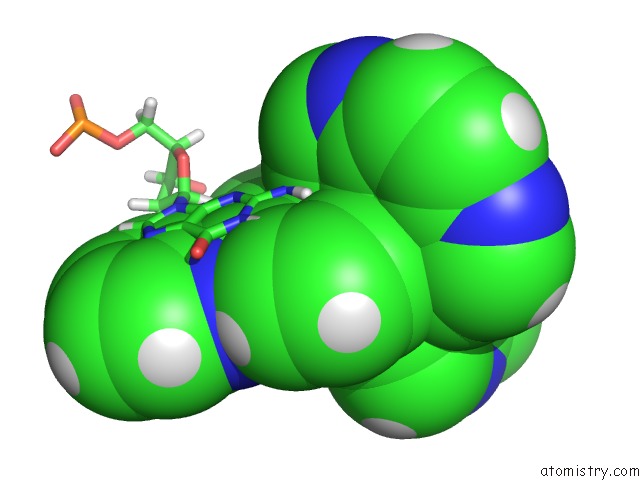
Mono view
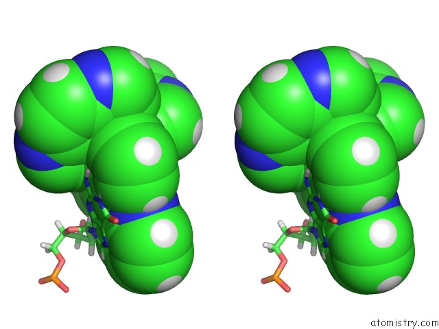
Stereo pair view
Mono view
Stereo pair view
A full contact list of Ruthenium with other atoms in the Ru binding
site number 1 of D(Tcggcgccga) with Lambda-[Ru(Tap)2(Dppz)]2+ Soaked in D2O within 5.0Å range:
|
Reference:
J.P.Hall,
F.E.Poynton,
P.M.Keane,
S.P.Gurung,
J.A.Brazier,
D.J.Cardin,
G.Winter,
T.Gunnlaugsson,
I.V.Sazanovich,
M.Towrie,
C.J.Cardin,
J.M.Kelly,
S.J.Quinn.
Monitoring One-Electron Photo-Oxidation of Guanine in Dna Crystals Using Ultrafast Infrared Spectroscopy. Nat Chem V. 7 961 2015.
ISSN: ESSN 1755-4349
PubMed: 26587711
DOI: 10.1038/NCHEM.2369
Page generated: Thu Oct 10 12:55:19 2024
ISSN: ESSN 1755-4349
PubMed: 26587711
DOI: 10.1038/NCHEM.2369
Last articles
Zn in 9MJ5Zn in 9HNW
Zn in 9G0L
Zn in 9FNE
Zn in 9DZN
Zn in 9E0I
Zn in 9D32
Zn in 9DAK
Zn in 8ZXC
Zn in 8ZUF