Ruthenium »
PDB 3m0b-4kgc »
4hhg »
Ruthenium in PDB 4hhg: Crystal Structure of the Pseudomonas Aeruginosa Azurin, RUH107NO YOH109
Protein crystallography data
The structure of Crystal Structure of the Pseudomonas Aeruginosa Azurin, RUH107NO YOH109, PDB code: 4hhg
was solved by
N.Herrera,
J.J.Warren,
H.B.Gray,
with X-Ray Crystallography technique. A brief refinement statistics is given in the table below:
| Resolution Low / High (Å) | 33.59 / 1.60 |
| Space group | I 2 2 2 |
| Cell size a, b, c (Å), α, β, γ (°) | 49.762, 67.176, 81.385, 90.00, 90.00, 90.00 |
| R / Rfree (%) | 23.4 / 26.1 |
Other elements in 4hhg:
The structure of Crystal Structure of the Pseudomonas Aeruginosa Azurin, RUH107NO YOH109 also contains other interesting chemical elements:
| Copper | (Cu) | 1 atom |
Ruthenium Binding Sites:
The binding sites of Ruthenium atom in the Crystal Structure of the Pseudomonas Aeruginosa Azurin, RUH107NO YOH109
(pdb code 4hhg). This binding sites where shown within
5.0 Angstroms radius around Ruthenium atom.
In total only one binding site of Ruthenium was determined in the Crystal Structure of the Pseudomonas Aeruginosa Azurin, RUH107NO YOH109, PDB code: 4hhg:
In total only one binding site of Ruthenium was determined in the Crystal Structure of the Pseudomonas Aeruginosa Azurin, RUH107NO YOH109, PDB code: 4hhg:
Ruthenium binding site 1 out of 1 in 4hhg
Go back to
Ruthenium binding site 1 out
of 1 in the Crystal Structure of the Pseudomonas Aeruginosa Azurin, RUH107NO YOH109
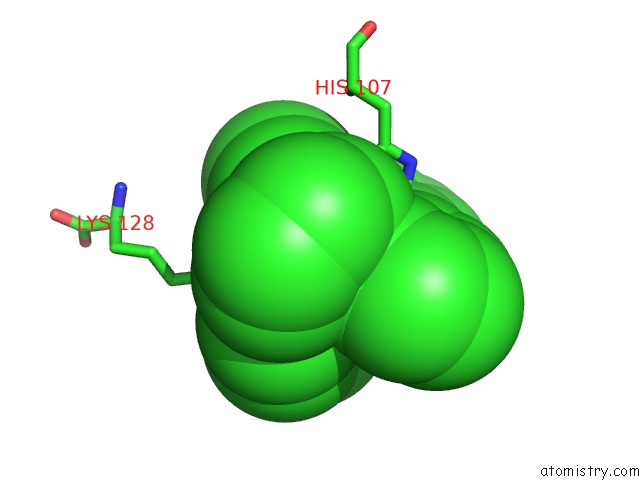
Mono view
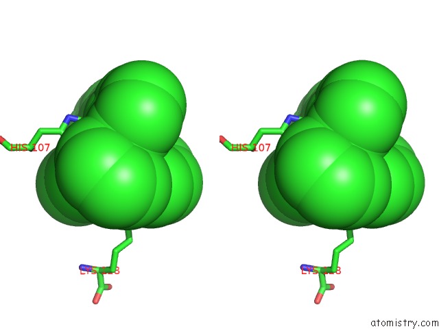
Stereo pair view
Mono view
Stereo pair view
A full contact list of Ruthenium with other atoms in the Ru binding
site number 1 of Crystal Structure of the Pseudomonas Aeruginosa Azurin, RUH107NO YOH109 within 5.0Å range:
|
Reference:
J.J.Warren,
N.Herrera,
M.G.Hill,
J.R.Winkler,
H.B.Gray.
Electron Flow Through Nitrotyrosinate in Pseudomonas Aeruginosa Azurin. J.Am.Chem.Soc. V. 135 11151 2013.
ISSN: ISSN 0002-7863
PubMed: 23859602
DOI: 10.1021/JA403734N
Page generated: Tue Aug 19 00:53:47 2025
ISSN: ISSN 0002-7863
PubMed: 23859602
DOI: 10.1021/JA403734N
Last articles
Sr in 3BNQSr in 2QJY
Sr in 2X53
Sr in 2SPT
Sr in 2XRM
Sr in 2WOH
Sr in 2RIO
Sr in 2PN4
Sr in 2QJK
Sr in 2QJP