Ruthenium »
PDB 3m0b-4kgc »
3qf8 »
Ruthenium in PDB 3qf8: X-Ray Crystal Structure of the Ruthenium Complex [Ru(Tap)2(Dppz)]2+ Bound to D(Tcggcgccga) at Medium Resolution
Protein crystallography data
The structure of X-Ray Crystal Structure of the Ruthenium Complex [Ru(Tap)2(Dppz)]2+ Bound to D(Tcggcgccga) at Medium Resolution, PDB code: 3qf8
was solved by
C.J.Cardin,
J.P.Hall,
with X-Ray Crystallography technique. A brief refinement statistics is given in the table below:
| Resolution Low / High (Å) | 13.96 / 1.73 |
| Space group | P 43 21 2 |
| Cell size a, b, c (Å), α, β, γ (°) | 42.239, 42.239, 39.379, 90.00, 90.00, 90.00 |
| R / Rfree (%) | 18.2 / 23.3 |
Other elements in 3qf8:
The structure of X-Ray Crystal Structure of the Ruthenium Complex [Ru(Tap)2(Dppz)]2+ Bound to D(Tcggcgccga) at Medium Resolution also contains other interesting chemical elements:
| Barium | (Ba) | 1 atom |
Ruthenium Binding Sites:
The binding sites of Ruthenium atom in the X-Ray Crystal Structure of the Ruthenium Complex [Ru(Tap)2(Dppz)]2+ Bound to D(Tcggcgccga) at Medium Resolution
(pdb code 3qf8). This binding sites where shown within
5.0 Angstroms radius around Ruthenium atom.
In total only one binding site of Ruthenium was determined in the X-Ray Crystal Structure of the Ruthenium Complex [Ru(Tap)2(Dppz)]2+ Bound to D(Tcggcgccga) at Medium Resolution, PDB code: 3qf8:
In total only one binding site of Ruthenium was determined in the X-Ray Crystal Structure of the Ruthenium Complex [Ru(Tap)2(Dppz)]2+ Bound to D(Tcggcgccga) at Medium Resolution, PDB code: 3qf8:
Ruthenium binding site 1 out of 1 in 3qf8
Go back to
Ruthenium binding site 1 out
of 1 in the X-Ray Crystal Structure of the Ruthenium Complex [Ru(Tap)2(Dppz)]2+ Bound to D(Tcggcgccga) at Medium Resolution
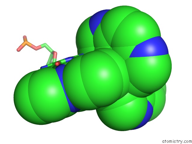
Mono view
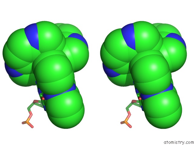
Stereo pair view
Mono view
Stereo pair view
A full contact list of Ruthenium with other atoms in the Ru binding
site number 1 of X-Ray Crystal Structure of the Ruthenium Complex [Ru(Tap)2(Dppz)]2+ Bound to D(Tcggcgccga) at Medium Resolution within 5.0Å range:
|
Reference:
J.P.Hall,
K.O'sullivan,
A.Naseer,
J.A.Smith,
J.M.Kelly,
C.J.Cardin.
Structure Determination of An Intercalating Ruthenium Dipyridophenazine Complex Which Kinks Dna By Semiintercalation of A Tetraazaphenanthrene Ligand. Proc.Natl.Acad.Sci.Usa V. 108 17610 2011.
ISSN: ISSN 0027-8424
PubMed: 21969542
DOI: 10.1073/PNAS.1108685108
Page generated: Thu Oct 10 12:49:34 2024
ISSN: ISSN 0027-8424
PubMed: 21969542
DOI: 10.1073/PNAS.1108685108
Last articles
Cl in 2VH0Cl in 2VG3
Cl in 2VGC
Cl in 2VG5
Cl in 2VG2
Cl in 2VFX
Cl in 2VF3
Cl in 2VFV
Cl in 2VFT
Cl in 2VFS