Ruthenium »
PDB 3m0b-4kgc »
3m0b »
Ruthenium in PDB 3m0b: Ru-Porphyrin Protein Scaffolds For Sensing O2
Protein crystallography data
The structure of Ru-Porphyrin Protein Scaffolds For Sensing O2, PDB code: 3m0b
was solved by
M.B.Winter,
E.J.Mclaurin,
S.Y.Reece,
C.Olea Jr.,
D.G.Nocera,
M.A.Marletta,
with X-Ray Crystallography technique. A brief refinement statistics is given in the table below:
| Resolution Low / High (Å) | 32.35 / 2.00 |
| Space group | P 61 2 2 |
| Cell size a, b, c (Å), α, β, γ (°) | 61.183, 61.183, 245.116, 90.00, 90.00, 120.00 |
| R / Rfree (%) | 20.4 / 22.5 |
Ruthenium Binding Sites:
The binding sites of Ruthenium atom in the Ru-Porphyrin Protein Scaffolds For Sensing O2
(pdb code 3m0b). This binding sites where shown within
5.0 Angstroms radius around Ruthenium atom.
In total only one binding site of Ruthenium was determined in the Ru-Porphyrin Protein Scaffolds For Sensing O2, PDB code: 3m0b:
In total only one binding site of Ruthenium was determined in the Ru-Porphyrin Protein Scaffolds For Sensing O2, PDB code: 3m0b:
Ruthenium binding site 1 out of 1 in 3m0b
Go back to
Ruthenium binding site 1 out
of 1 in the Ru-Porphyrin Protein Scaffolds For Sensing O2
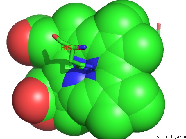
Mono view
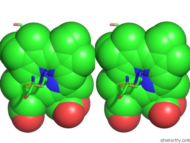
Stereo pair view
Mono view
Stereo pair view
A full contact list of Ruthenium with other atoms in the Ru binding
site number 1 of Ru-Porphyrin Protein Scaffolds For Sensing O2 within 5.0Å range:
|
Reference:
M.B.Winter,
E.J.Mclaurin,
S.Y.Reece,
C.Olea,
D.G.Nocera,
M.A.Marletta.
Ru-Porphyrin Protein Scaffolds For Sensing O2. J.Am.Chem.Soc. V. 132 5582 2010.
ISSN: ISSN 0002-7863
PubMed: 20373741
DOI: 10.1021/JA101527R
Page generated: Thu Oct 10 12:48:50 2024
ISSN: ISSN 0002-7863
PubMed: 20373741
DOI: 10.1021/JA101527R
Last articles
Zn in 9MJ5Zn in 9HNW
Zn in 9G0L
Zn in 9FNE
Zn in 9DZN
Zn in 9E0I
Zn in 9D32
Zn in 9DAK
Zn in 8ZXC
Zn in 8ZUF